|
|
|
|
 Description
|
|
Compounds
|
||||||||||||||||||||
Chains, Units
Summary Information (see also Sequences/Alignments below) |
Ligands, Modified Residues, Ions (0, 0)| (no "Ligand,Modified Residues,Ions" information available for 1PHS) |
Sites (0, 0)| (no "Site" information available for 1PHS) |
SS Bonds (0, 0)| (no "SS Bond" information available for 1PHS) |
Cis Peptide Bonds (0, 0)| (no "Cis Peptide Bond" information available for 1PHS) |
SAPs(SNPs)/Variants (0, 0)| (no "SAP(SNP)/Variant" information available for 1PHS) |
PROSITE Motifs (0, 0)| (no "PROSITE Motif" information available for 1PHS) |
Exons (0, 0)| (no "Exon" information available for 1PHS) |
Sequences/Alignments
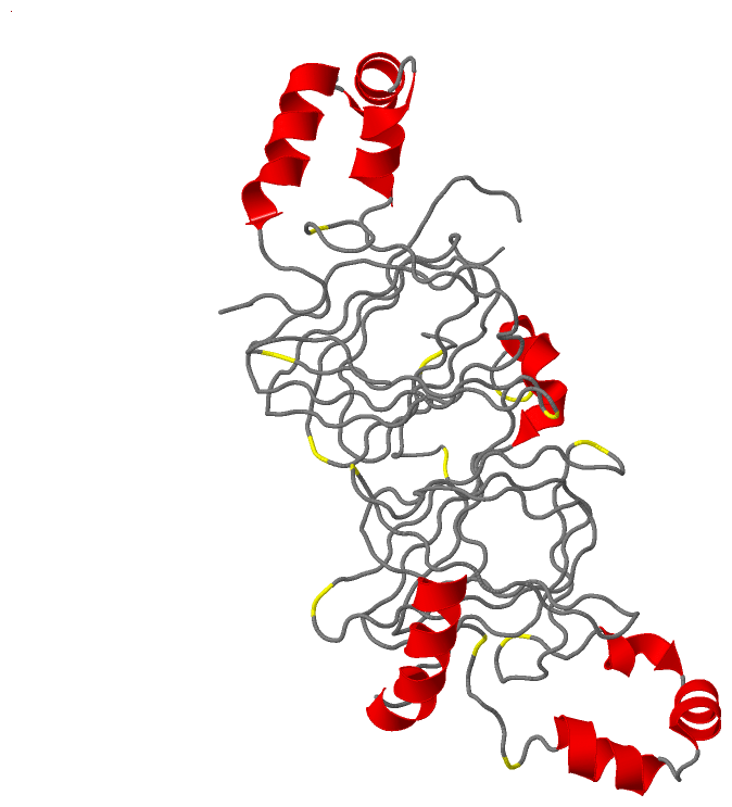
Asymmetric/Biological UnitChain A from PDB Type:PROTEIN Length:364 aligned with PHSB_PHAVU | P02853 from UniProtKB/Swiss-Prot Length:421 Alignment length:371 44 54 64 74 84 94 104 114 124 134 144 154 164 174 184 194 204 214 224 234 244 254 264 274 284 294 304 314 324 334 344 354 364 374 384 394 404 PHSB_PHAVU 35 DNPFYFNSDNSWNTLFKNQYGHIRVLQRFDQQSKRLQNLEDYRLVEFRSKPETLLLPQQADAELLLVVRSGSAILVLVKPDDRREYFFLTSDNPIFSDHQKIPAGTIFYLVNPDPKEDLRIIQLAMPVNNPQIHEFFLSSTEAQQSYLQEFSKHILEASFNSKFEEINRVLFEEEGQQEGVIVNIDSEQIKELSKHAKSSSRKSLSKQDNTIGNEFGNLTERTDNSLNVLISSIEMEEGALFVPHYYSKAIVILVVNEGEAHVELVGPKGNKETLEYESYRAELSKDDVFVIPAAYPVAIKATSNVNFTGFGINANNNNRNLLAGKTDNVISSIGRALDGKDVLGLTFSGSGDEVMKLINKQSGSYFVDAH 405 SCOP domains d1phsa1 A:11-212 Seed storage 7S protein -------d1phsa2 A:220-381 Seed storage 7S protein SCOP domains CATH domains ----------------------------------------------------------------------------------------------------------------------------------------------------------------------------------------------------------------------------------------------------------------------------------------------------------------------------------------------------------------------------------- CATH domains Pfam domains ----------------------------------------------------------------------------------------------------------------------------------------------------------------------------------------------------------------------------------------------------------------------------------------------------------------------------------------------------------------------------------- Pfam domains SAPs(SNPs) ----------------------------------------------------------------------------------------------------------------------------------------------------------------------------------------------------------------------------------------------------------------------------------------------------------------------------------------------------------------------------------- SAPs(SNPs) PROSITE ----------------------------------------------------------------------------------------------------------------------------------------------------------------------------------------------------------------------------------------------------------------------------------------------------------------------------------------------------------------------------------- PROSITE Transcript ----------------------------------------------------------------------------------------------------------------------------------------------------------------------------------------------------------------------------------------------------------------------------------------------------------------------------------------------------------------------------------- Transcript 1phs A 11 DNPFYFNSDNSWNTLFKNQYGHIRVLQRFDQQSKRLQNLEDYRLVEFRSKPETLLLPQQADAELLLVVRSGSAILVLVKPDDRREYFFLTSDNPIFSDHQKIPAGTIFYLVNPDPKEDLRIIQLAMPVNNPQIHEFFLSSTEAQQSYLQEFSKHILEASFNSKFEEINRVLFEEEGQQEGVIVNIDSEQIKELSKHAKSSSR-------NTIGNEFGNLTERTDNSLNVLISSIEMEEGALFVPHYYSKAIVILVVNEGEAHVELVGPKGNKETLEYESYRAELSKDDVFVIPAAYPVAIKATSNVNFTGFGINANNNNRNLLAGKTDNVISSIGRALDGKDVLGLTFSGSGDEVMKLINKQSGSYFVDAH 381 20 30 40 50 60 70 80 90 100 110 120 130 140 150 160 170 180 190 200 210 | 220 230 240 250 260 270 280 290 300 310 320 330 340 350 360 370 380 212 220
|
||||||||||||||||||||
SCOP Domains (1, 2)
Asymmetric/Biological Unit
 |
CATH Domains (0, 0)| (no "CATH Domain" information available for 1PHS) |
Pfam Domains (0, 0)| (no "Pfam Domain" information available for 1PHS) |
Gene Ontology (3, 3)|
Asymmetric/Biological Unit(hide GO term definitions) Chain A (PHSB_PHAVU | P02853)
|
||||||||||||||||||||||||||||||
Interactive Views
|
||||||||||||||||||||||||||||||||||||||||||||||||||||||||||||||||||||||||||||||||||||||||||||||||||||||||||||||||||||
Still Images
|
||||||||||||||||
Databases
|
||||||||||||||||||||||||||||||||||||||||||||||||||||||||||||||||||||||||||||||||||||||||||||||||||||||||||||||||||||||||||||||||||||||||||||||||||||||||||||||||
Analysis Tools
|
|||||||||||||||||||||||||||||||||||||||||||||||||||||||||||||
Entries Sharing at Least One Protein Chain (UniProt ID)
Related Entries Specified in the PDB File
|
|