|
|
|
|
 Description
|
|
Compounds
|
||||||||||||||||||||||||
Chains, Units
Summary Information (see also Sequences/Alignments below) |
Ligands, Modified Residues, Ions (1, 1)
Asymmetric/Biological Unit (1, 1)
|
Sites (1, 1)
Asymmetric Unit (1, 1)
|
SS Bonds (0, 0)| (no "SS Bond" information available for 1PHR) |
Cis Peptide Bonds (0, 0)| (no "Cis Peptide Bond" information available for 1PHR) |
SAPs(SNPs)/Variants (0, 0)| (no "SAP(SNP)/Variant" information available for 1PHR) |
PROSITE Motifs (0, 0)| (no "PROSITE Motif" information available for 1PHR) |
Exons (6, 6)
Asymmetric/Biological Unit (6, 6)
|
||||||||||||||||||||||||||||||||||||||||||||||||||||||||||||||||||||||||||||||||||||||||||||||||
Sequences/Alignments
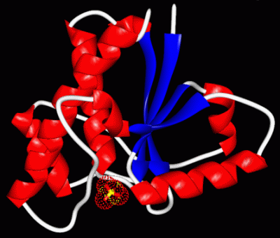
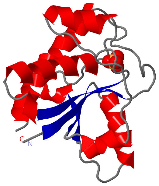
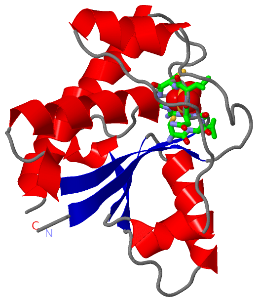
Asymmetric/Biological UnitChain A from PDB Type:PROTEIN Length:154 aligned with PPAC_BOVIN | P11064 from UniProtKB/Swiss-Prot Length:158 Alignment length:154 14 24 34 44 54 64 74 84 94 104 114 124 134 144 154 PPAC_BOVIN 5 VTKSVLFVCLGNICRSPIAEAVFRKLVTDQNISDNWVIDSGAVSDWNVGRSPDPRAVSCLRNHGINTAHKARQVTKEDFVTFDYILCMDESNLRDLNRKSNQVKNCRAKIELLGSYDPQKQLIIEDPYYGNDADFETVYQQCVRCCRAFLEKVR 158 SCOP domains d1phra_ A: Tyrosine phosphatase SCOP domains CATH domains 1phrA00 A:4-157 [code=3.40.50.270, no name defined] CATH domains Pfam domains ----LMWPc-1phrA01 A:8-153 ---- Pfam domains SAPs(SNPs) ---------------------------------------------------------------------------------------------------------------------------------------------------------- SAPs(SNPs) PROSITE ---------------------------------------------------------------------------------------------------------------------------------------------------------- PROSITE Transcript 1 (1) Exon 1.1 ------------------------Exon 1.4 PDB: A:39-76 UniProt: 40-77 Exon 1.5 PDB: A:77-9-----------------------------------Exon 1.7 PDB: A:133-157 Transcript 1 (1) Transcript 1 (2) ----------Exon 1.2 PDB: A:14-38 ----------------------------------------------------------Exon 1.6 PDB: A:97-132 ------------------------- Transcript 1 (2) 1phr A 4 VTKSVLFVCLGNICRSPIAEAVFRKLVTDQNISDNWVIDSGAVSDWNVGRSPDPRAVSCLRNHGINTAHKARQVTKEDFVTFDYILCMDESNLRDLNRKSNQVKNCRAKIELLGSYDPQKQLIIEDPYYGNDADFETVYQQCVRCCRAFLEKVR 157 13 23 33 43 53 63 73 83 93 103 113 123 133 143 153
|
||||||||||||||||||||
SCOP Domains (1, 1)
Asymmetric/Biological Unit
 |
CATH Domains (1, 1)
Asymmetric/Biological Unit
|
Pfam Domains (1, 1)
Asymmetric/Biological Unit
|
Gene Ontology (8, 8)|
Asymmetric/Biological Unit(hide GO term definitions) Chain A (PPAC_BOVIN | P11064)
|
||||||||||||||||||||||||||||||||||||||||||||||||||||||||||||||||||
Interactive Views
|
||||||||||||||||||||||||||||||||||||||||||||||||||||||||||||||||||||||||||||||||||||||||||||||||||||||||||||||||||||||
Still Images
|
||||||||||||||||||||||||||||||||||||||||||||||||||||||||||||||
Databases
|
||||||||||||||||||||||||||||||||||||||||||||||||||||||||||||||||||||||||||||||||||||||||||||||||||||||||||||||||||||||||||||||||||||||||||||||||||||||||||||||||
Analysis Tools
|
|||||||||||||||||||||||||||||||||||||||||||||||||||||||||||||
Entries Sharing at Least One Protein Chain (UniProt ID)
Related Entries Specified in the PDB File
|
|