Show PDB file:   
         Plain Text   HTML   (compressed file size)
QuickSearch:   
by PDB,NDB,UniProt,PROSITE Code or Search Term(s)  
(-)Asymmetric Unit
(-)Biological Unit 1
(-)Biological Unit 2
collapse expand < >
Image Asymmetric Unit
Asymmetric Unit  (Jmol Viewer)
Image Biological Unit 1
Biological Unit 1  (Jmol Viewer)
Image Biological Unit 2
Biological Unit 2  (Jmol Viewer)

(-) Description

Title :  CRYSTAL STRUCTURE OF OXIDIZED MERP FROM RALSTONIA METALLIDURANS CH34
 
Authors :  L. Serre, E. Rossy, E. Pebay-Peyroula, C. Cohen-Addad, J. Coves
Date :  19 Mar 03  (Deposition) - 18 May 04  (Release) - 24 Feb 09  (Revision)
Method :  X-RAY DIFFRACTION
Resolution :  2.00
Chains :  Asym. Unit :  A,B
Biol. Unit 1:  A  (1x)
Biol. Unit 2:  B  (1x)
Keywords :  Mercury Resistance, Metal Binding Protein, Perisplasm, Structural Genomics (Keyword Search: [Gene Ontology, PubMed, Web (Google))
 
Reference :  L. Serre, E. Rossy, E. Pebay-Peyroula, C. Cohen-Addad, J. Coves
Crystal Structure Of The Oxidized Form Of The Periplasmic Mercury-Binding Protein Merp From Ralstonia Metallidurans Ch34
J. Mol. Biol. V. 339 161 2004
PubMed-ID: 15123428  |  Reference-DOI: 10.1016/J.JMB.2004.03.022
(for further references see the PDB file header)

(-) Compounds

Molecule 1 - HYPOTHETICAL PROTEIN MERP
    ChainsA, B
    EngineeredYES
    Expression SystemESCHERICHIA COLI BL21(DE3)
    Expression System PlasmidPET-3A
    Expression System StrainBL21 (DE3)
    Expression System Taxid469008
    Expression System Vector TypePLASMID
    FragmentRESIDUES 1-72
    GeneMERP
    Organism ScientificCUPRIAVIDUS METALLIDURANS
    Organism Taxid119219
    SynonymMERP

 Structural Features

(-) Chains, Units

  12
Asymmetric Unit AB
Biological Unit 1 (1x)A 
Biological Unit 2 (1x) B

Summary Information (see also Sequences/Alignments below)

(-) Ligands, Modified Residues, Ions  (0, 0)

(no "Ligand,Modified Residues,Ions" information available for 1OSD)

(-) Sites  (0, 0)

(no "Site" information available for 1OSD)
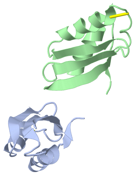
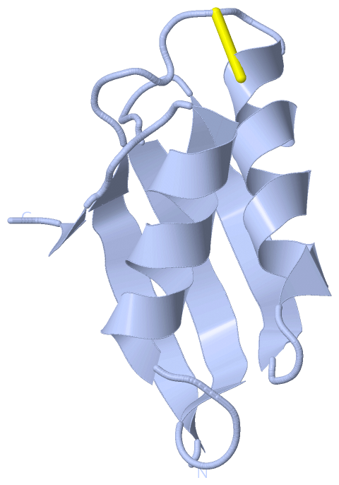
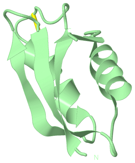
(-) SS Bonds  (2, 2)

Asymmetric Unit
No.Residues
1A:14 -A:17
2B:14 -B:17

(-) Cis Peptide Bonds  (0, 0)

(no "Cis Peptide Bond" information available for 1OSD)

 Sequence-Structure Mapping

(-) SAPs(SNPs)/Variants  (0, 0)

(no "SAP(SNP)/Variant" information available for 1OSD)

(-) PROSITE Motifs  (0, 0)

(no "PROSITE Motif" information available for 1OSD)

(-) Exons   (0, 0)

(no "Exon" information available for 1OSD)

(-) Sequences/Alignments

Asymmetric Unit
   Reformat: Number of residues per line =  ('0' or empty: single-line sequence representation)
  Number of residues per labelling interval =   
  UniProt sequence: complete  aligned part    
   Show mapping: SCOP domains CATH domains Pfam domains Secondary structure (by author)
SAPs(SNPs) PROSITE motifs Exons
(details for a mapped element are shown in a popup box when the mouse pointer rests over it)
Chain A from PDB  Type:PROTEIN  Length:72
 aligned with Q5NUU9_CUPMC | Q5NUU9 from UniProtKB/TrEMBL  Length:87

    Alignment length:72
                                    25        35        45        55        65        75        85  
          Q5NUU9_CUPMC   16 ATQTVTLSVPGMTCSACPITVKKAISKVEGVSKVDVTFETRQAVVTFDDAKTSVQKLTKATADAGYPSSVKQ 87
               SCOP domains d1osda_ A: Mercuric ion binding protein MerP                             SCOP domains
               CATH domains 1osdA00 A:1-72  [code=3.30.70.100, no name defined]                      CATH domains
               Pfam domains ------------------------------------------------------------------------ Pfam domains
         Sec.struct. author .eeeeeee.......hhhhhhhhhhhh...eeeeeee....eeeeeee....hhhhhhhhhhhh....ee.. Sec.struct. author
                 SAPs(SNPs) ------------------------------------------------------------------------ SAPs(SNPs)
                    PROSITE ------------------------------------------------------------------------ PROSITE
                 Transcript ------------------------------------------------------------------------ Transcript
                  1osd A  1 ATQTVTLSVPGMTCSACPITVKKAISKVEGVSKVDVTFETRQAVVTFDDAKTSVQKLTKATADAGYPSSVKQ 72
                                    10        20        30        40        50        60        70  

Chain B from PDB  Type:PROTEIN  Length:72
 aligned with Q5NUU9_CUPMC | Q5NUU9 from UniProtKB/TrEMBL  Length:87

    Alignment length:72
                                    25        35        45        55        65        75        85  
          Q5NUU9_CUPMC   16 ATQTVTLSVPGMTCSACPITVKKAISKVEGVSKVDVTFETRQAVVTFDDAKTSVQKLTKATADAGYPSSVKQ 87
               SCOP domains d1osdb_ B: Mercuric ion binding protein MerP                             SCOP domains
               CATH domains 1osdB00 B:1-72  [code=3.30.70.100, no name defined]                      CATH domains
               Pfam domains ------------------------------------------------------------------------ Pfam domains
         Sec.struct. author .eeeeeee.......hhhhhhhhhhhh...eeeeeee....eeeeeee....hhhhhhhhhhhh....ee.. Sec.struct. author
                 SAPs(SNPs) ------------------------------------------------------------------------ SAPs(SNPs)
                    PROSITE ------------------------------------------------------------------------ PROSITE
                 Transcript ------------------------------------------------------------------------ Transcript
                  1osd B  1 ATQTVTLSVPGMTCSACPITVKKAISKVEGVSKVDVTFETRQAVVTFDDAKTSVQKLTKATADAGYPSSVKQ 72
                                    10        20        30        40        50        60        70  

   Legend:   → Mismatch (orange background)
  - → Gap (green background, '-', border residues have a numbering label)
    → Modified Residue (blue background, lower-case, 'x' indicates undefined single-letter code, labelled with number + name)
  x → Chemical Group (purple background, 'x', labelled with number + name, e.g. ACE or NH2)
  extra numbering lines below/above indicate numbering irregularities and modified residue names etc., number ends below/above '|'

 Classification and Annotation

(-) SCOP Domains  (1, 2)

Asymmetric Unit

(-) CATH Domains  (1, 2)

Asymmetric Unit
(-)
Class: Alpha Beta (26913)

(-) Pfam Domains  (0, 0)

(no "Pfam Domain" information available for 1OSD)

(-) Gene Ontology  (7, 7)

Asymmetric Unit(hide GO term definitions)
Chain A,B   (Q5NUU9_CUPMC | Q5NUU9)
molecular function
    GO:0045340    mercury ion binding    Interacting selectively and non-covalently with mercury (Hg) ions.
    GO:0015097    mercury ion transmembrane transporter activity    Enables the transfer of mercury (Hg) ions from one side of a membrane to the other.
    GO:0046872    metal ion binding    Interacting selectively and non-covalently with any metal ion.
biological process
    GO:0015694    mercury ion transport    The directed movement of mercury (Hg) ions into, out of or within a cell, or between cells, by means of some agent such as a transporter or pore.
    GO:0030001    metal ion transport    The directed movement of metal ions, any metal ion with an electric charge, into, out of or within a cell, or between cells, by means of some agent such as a transporter or pore.
    GO:0046689    response to mercury ion    Any process that results in a change in state or activity of a cell or an organism (in terms of movement, secretion, enzyme production, gene expression, etc.) as a result of a mercury ion stimulus.
cellular component
    GO:0042597    periplasmic space    The region between the inner (cytoplasmic) and outer membrane (Gram-negative Bacteria) or cytoplasmic membrane and cell wall (Fungi and Gram-positive Bacteria).

 Visualization

(-) Interactive Views

Asymmetric Unit
  Complete Structure
    Jena3D(integrated viewing of ligand, site, SAP, PROSITE, SCOP information)
    WebMol | AstexViewer[tm]@PDBe
(Java Applets, require no local installation except for Java; loading may be slow)
    STRAP
(Java WebStart application, automatic local installation, requires Java; full application with system access!)
    RasMol
(require local installation)
    Molscript (VRML)
(requires installation of a VRML viewer; select preferred view via VRML and generate a mono or stereo PDF format file)
 
  Ligands, Modified Residues, Ions
(no "Ligands, Modified Residues, Ions" information available for 1osd)
 
  Sites
(no "Sites" information available for 1osd)
 
  Cis Peptide Bonds
(no "Cis Peptide Bonds" information available for 1osd)
 
Biological Units
  Complete Structure
    Biological Unit 1  [ Jena3D ]
    Biological Unit 2  [ Jena3D ]

(-) Still Images

Jmol
  protein: cartoon or spacefill or dots and stick; nucleic acid: cartoon and stick; ligands: spacefill; active site: stick
Molscript
  protein, nucleic acid: cartoon; ligands: spacefill; active site: ball and stick

 Databases and Analysis Tools

(-) Databases

Access by PDB/NDB ID
  1osd
    Family and Domain InformationProDom | SYSTERS
    General Structural InformationGlycoscienceDB | MMDB | NDB | OCA | PDB | PDBe | PDBj | PDBsum | PDBWiki | PQS | PROTEOPEDIA
    Orientation in MembranesOPM
    Protein SurfaceSURFACE
    Secondary StructureDSSP (structure derived) | HSSP (homology derived)
    Structural GenomicsGeneCensus
    Structural NeighboursCE | VAST
    Structure ClassificationCATH | Dali | SCOP
    Validation and Original DataBMRB Data View | BMRB Restraints Grid | EDS | PROCHECK | RECOORD | WHAT_CHECK
 
Access by UniProt ID/Accession number
  Q5NUU9_CUPMC | Q5NUU9
    Comparative Protein Structure ModelsModBase
    Genomic InformationEnsembl
    Protein-protein InteractionDIP
    Sequence, Family and Domain InformationInterPro | Pfam | SMART | UniProtKB/TrEMBL
 
Access by Enzyme Classificator   (EC Number)
  (no 'Enzyme Classificator' available)
    General Enzyme InformationBRENDA | EC-PDB | Enzyme | IntEnz
    PathwayKEGG | MetaCyc
 
Access by Disease Identifier   (MIM ID)
  (no 'MIM ID' available)
    Disease InformationOMIM
 
Access by GenAge ID
  (no 'GenAge ID' available)
    Age Related InformationGenAge

(-) Analysis Tools

Access by PDB/NDB ID
    Domain InformationXDom
    Interatomic Contacts of Structural UnitsCSU
    Ligand-protein ContactsLPC
    Protein CavitiescastP
    Sequence and Secondary StructurePDBCartoon
    Structure AlignmentSTRAP(Java WebStart application, automatic local installation, requires Java; full application with system access!)
    Structure and Sequence BrowserSTING
 
Access by UniProt ID/Accession number
  Q5NUU9_CUPMC | Q5NUU9
    Protein Disorder PredictionDisEMBL | FoldIndex | GLOBPLOT (for more information see DisProt)

 Related Entries

(-) Entries Sharing at Least One Protein Chain (UniProt ID)

(no "Entries Sharing at Least One Protein Chain" available for 1OSD)

(-) Related Entries Specified in the PDB File

1afi SHIGELLA FLEXNERI REDUCED MERP
1afj SHIGELLA FLEXNERI MERCURY BOUND MERP
2hqi SHIGELLA FLEXNERI OXIDIZED MERP