|
|
|
|
 Description
|
|
Compounds
|
||||||||||||||||||||||||||||||||||||||||||||
Chains, Units
Summary Information (see also Sequences/Alignments below) |
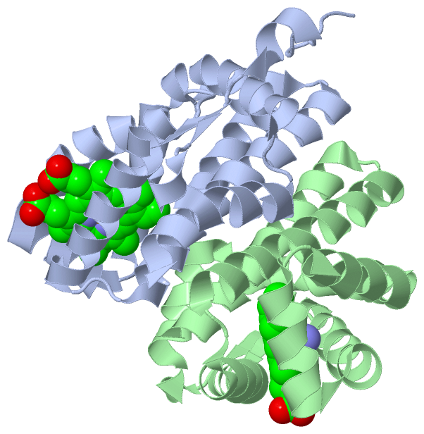
Ligands, Modified Residues, Ions (2, 4)| Asymmetric/Biological Unit (2, 4) |
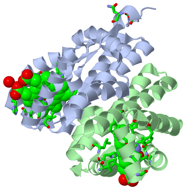
Sites (4, 4)
Asymmetric Unit (4, 4)
|
SS Bonds (0, 0)| (no "SS Bond" information available for 1OR4) |
Cis Peptide Bonds (1, 1)
Asymmetric/Biological Unit
|
||||||||
SAPs(SNPs)/Variants (0, 0)| (no "SAP(SNP)/Variant" information available for 1OR4) |
PROSITE Motifs (0, 0)| (no "PROSITE Motif" information available for 1OR4) |
Exons (0, 0)| (no "Exon" information available for 1OR4) |
Sequences/Alignments
Asymmetric/Biological UnitChain A from PDB Type:PROTEIN Length:169 aligned with HEMAT_BACSU | O07621 from UniProtKB/Swiss-Prot Length:432 Alignment length:169 19 29 39 49 59 69 79 89 99 109 119 129 139 149 159 169 HEMAT_BACSU 10 ETAYFSDSNGQQKNRIQLTNKHADVKKQLKMVRLGDAELYVLEQLQPLIQENIVNIVDAFYKNLDHESSLMDIINDHSSVDRLKQTLKRHIQEMFAGVIDDEFIEKRNRIASIHLRIGLLPKWYMGAFQELLLSMIDIYEASITNQQELLKAIKATTKILNLEQQLVLE 178 SCOP domains d1or4a_ A: Heme-based aerotactic transducer HemAT, sensor domain SCOP domains CATH domains 1or4A00 A:10-178 Globins CATH domains Pfam domains ------------------------------------------------------------------------------------------------------------------------------------------------------------------------- Pfam domains SAPs(SNPs) ------------------------------------------------------------------------------------------------------------------------------------------------------------------------- SAPs(SNPs) PROSITE ------------------------------------------------------------------------------------------------------------------------------------------------------------------------- PROSITE Transcript ------------------------------------------------------------------------------------------------------------------------------------------------------------------------- Transcript 1or4 A 10 ETAYFSDSNGQQKNRIQLTNKHADVKKQLKMVRLGDAELYVLEQLQPLIQENIVNIVDAFYKNLDHESSLMDIINDHSSVDRLKQTLKRHIQEMFAGVIDDEFIEKRNRIASIHLRIGLLPKWYMGAFQELLLSMIDIYEASITNQQELLKAIKATTKILNLEQQLVLE 178 19 29 39 49 59 69 79 89 99 109 119 129 139 149 159 169 Chain B from PDB Type:PROTEIN Length:158 aligned with HEMAT_BACSU | O07621 from UniProtKB/Swiss-Prot Length:432 Alignment length:158 30 40 50 60 70 80 90 100 110 120 130 140 150 160 170 HEMAT_BACSU 21 QKNRIQLTNKHADVKKQLKMVRLGDAELYVLEQLQPLIQENIVNIVDAFYKNLDHESSLMDIINDHSSVDRLKQTLKRHIQEMFAGVIDDEFIEKRNRIASIHLRIGLLPKWYMGAFQELLLSMIDIYEASITNQQELLKAIKATTKILNLEQQLVLE 178 SCOP domains d1or4b_ B: Heme-based aerotactic transducer HemAT, sensor domain SCOP domains CATH domains 1or4B00 B:21-178 Globins CATH domains Pfam domains -------------------------------------------------------------------------------------------------------------------------------------------------------------- Pfam domains SAPs(SNPs) -------------------------------------------------------------------------------------------------------------------------------------------------------------- SAPs(SNPs) PROSITE -------------------------------------------------------------------------------------------------------------------------------------------------------------- PROSITE Transcript -------------------------------------------------------------------------------------------------------------------------------------------------------------- Transcript 1or4 B 21 QKNRIQLTNKHADVKKQLKMVRLGDAELYVLEQLQPLIQENIVNIVDAFYKNLDHESSLMDIINDHSSVDRLKQTLKRHIQEMFAGVIDDEFIEKRNRIASIHLRIGLLPKWYMGAFQELLLSMIDIYEASITNQQELLKAIKATTKILNLEQQLVLE 178 30 40 50 60 70 80 90 100 110 120 130 140 150 160 170
|
||||||||||||||||||||
SCOP Domains (1, 2)
Asymmetric/Biological Unit
 |
CATH Domains (1, 2)
Asymmetric/Biological Unit
|
Pfam Domains (0, 0)| (no "Pfam Domain" information available for 1OR4) |
Gene Ontology (8, 8)|
Asymmetric/Biological Unit(hide GO term definitions) Chain A,B (HEMAT_BACSU | O07621)
|
||||||||||||||||||||||||||||||||||||||||||||||||||||||||||||||||||
Interactive Views
|
|||||||||||||||||||||||||||||||||||||||||||||||||||||||||||||||||||||||||||||||||||||||||||||||||||||||||||||||||||||||||||||||||||||||||||||||||||
Still Images
|
||||||||||||||||
Databases
|
||||||||||||||||||||||||||||||||||||||||||||||||||||||||||||||||||||||||||||||||||||||||||||||||||||||||||||||||||||||||||||||||||||||||||||||||||||||||||||||||
Analysis Tools
|
|||||||||||||||||||||||||||||||||||||||||||||||||||||||||||||
Entries Sharing at Least One Protein Chain (UniProt ID)
Related Entries Specified in the PDB File
|
|