| molecular function |
|---|
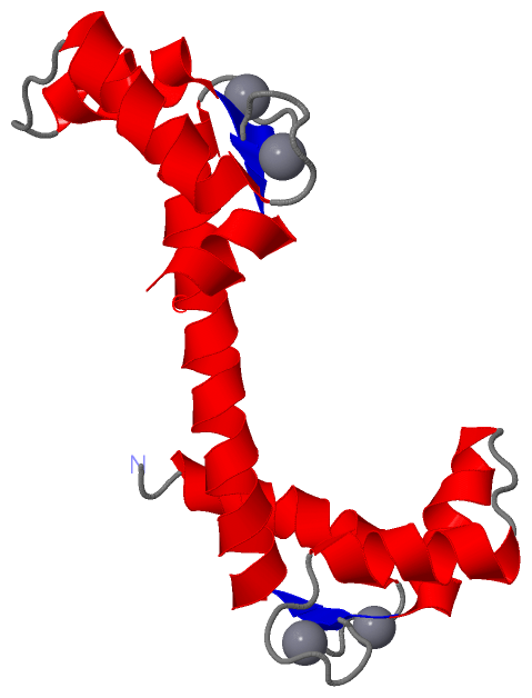
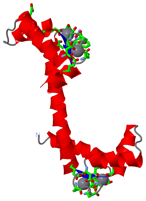
| | GO:0005509 | | calcium ion binding | | Interacting selectively and non-covalently with calcium ions (Ca2+). |
| | GO:0046872 | | metal ion binding | | Interacting selectively and non-covalently with any metal ion. |
| | GO:0005515 | | protein binding | | Interacting selectively and non-covalently with any protein or protein complex (a complex of two or more proteins that may include other nonprotein molecules). |
| biological process |
|---|
| | GO:0043277 | | apoptotic cell clearance | | The recognition and removal of an apoptotic cell by a neighboring cell or by a phagocyte. |
| | GO:0016477 | | cell migration | | The controlled self-propelled movement of a cell from one site to a destination guided by molecular cues. Cell migration is a central process in the development and maintenance of multicellular organisms. |
| | GO:0009792 | | embryo development ending in birth or egg hatching | | The process whose specific outcome is the progression of an embryo over time, from zygote formation until the end of the embryonic life stage. The end of the embryonic life stage is organism-specific and may be somewhat arbitrary; for mammals it is usually considered to be birth, for insects the hatching of the first instar larva from the eggshell. |
| | GO:0051296 | | establishment of meiotic spindle orientation | | Any process that set the alignment of meiotic spindle relative to other cellular structures. |
| | GO:0042981 | | regulation of apoptotic process | | Any process that modulates the occurrence or rate of cell death by apoptotic process. |
| | GO:0051726 | | regulation of cell cycle | | Any process that modulates the rate or extent of progression through the cell cycle. |
| | GO:0032880 | | regulation of protein localization | | Any process that modulates the frequency, rate or extent of any process in which a protein is transported to, or maintained in, a specific location. |
| cellular component |
|---|
| | GO:0071944 | | cell periphery | | The part of a cell encompassing the cell cortex, the plasma membrane, and any external encapsulating structures. |
| | GO:0005813 | | centrosome | | A structure comprised of a core structure (in most organisms, a pair of centrioles) and peripheral material from which a microtubule-based structure, such as a spindle apparatus, is organized. Centrosomes occur close to the nucleus during interphase in many eukaryotic cells, though in animal cells it changes continually during the cell-division cycle. |
| | GO:0072686 | | mitotic spindle | | A spindle that forms as part of mitosis. Mitotic and meiotic spindles contain distinctive complements of proteins associated with microtubules. |
| | GO:0031965 | | nuclear membrane | | Either of the lipid bilayers that surround the nucleus and form the nuclear envelope; excludes the intermembrane space. |
 Description