|
|
|
|
 Description
|
|
Compounds
|
||||||||||||||||||||||||||||
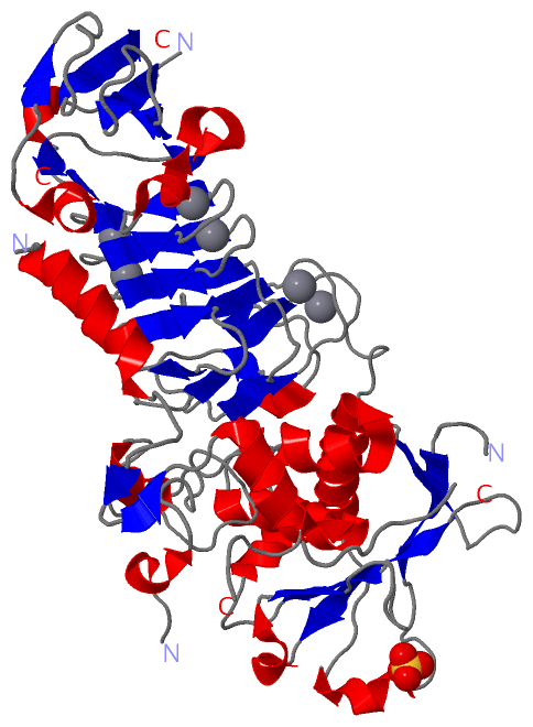
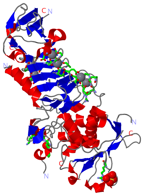
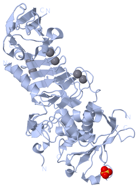
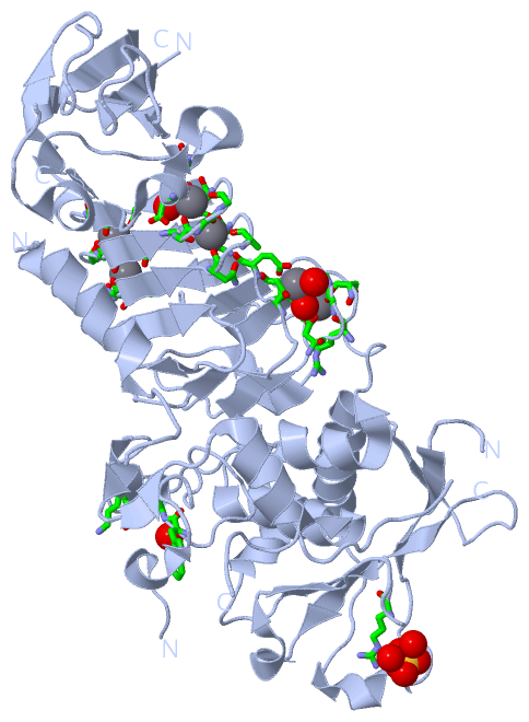
Chains, Units
Summary Information (see also Sequences/Alignments below) |
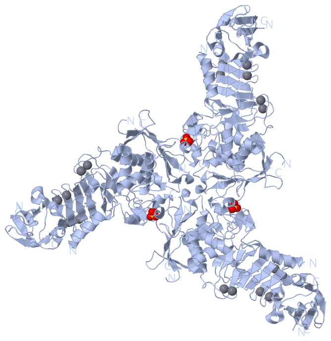
Ligands, Modified Residues, Ions (2, 7)| Asymmetric Unit (2, 7) Biological Unit 1 (1, 1) Biological Unit 2 (1, 3) |
Sites (7, 7)
Asymmetric Unit (7, 7)
|
SS Bonds (0, 0)| (no "SS Bond" information available for 1OM8) |
Cis Peptide Bonds (0, 0)| (no "Cis Peptide Bond" information available for 1OM8) |
SAPs(SNPs)/Variants (0, 0)| (no "SAP(SNP)/Variant" information available for 1OM8) |
PROSITE Motifs (0, 0)| (no "PROSITE Motif" information available for 1OM8) |
Exons (0, 0)| (no "Exon" information available for 1OM8) |
Sequences/Alignments
Asymmetric UnitChain A from PDB Type:PROTEIN Length:452 aligned with O69771_9PSED | O69771 from UniProtKB/TrEMBL Length:480 Alignment length:461 29 39 49 59 69 79 89 99 109 119 129 139 149 159 169 179 189 199 209 219 229 239 249 259 269 279 289 299 309 319 329 339 349 359 369 379 389 399 409 419 429 439 449 459 469 479 O69771_9PSED 20 GTSSAFTQIDNFSHFYDRGEHLVNGKPSFTVDQVADQLTRSGASWHDLNNDGVINLTYTFLTAPPVGYASRGLGTFSQFSALQKEQAKLSLESWADVAKVTFTEGPAARGDDGHMTFANFSASNGGAAFAYLPNSSRKGESWYLINKDYQVNKTPGEGNYGRQTLTHEIGHTLGLSHPGDYNAGNGNPTYRDAVYAEDTRAYSVMSYWSEKNTGQVFTKTGEGAYASAPLLDDIAAVQKLYGANLETRADDTVYGFNSTADRDFYSATSSTDKLIFSVWDGGGNDTLDFSGFSQNQKINLTAGSFSDVGGMTGNVSIAQGVTIENAIGGSGNDLLIGNDAANVLKGGAGNDIIYGGGGADVLWGGTGSDTFVFGAVSDSTPKAADIIKDFQSGFDKIDLTAITKLGGLNFVDAFTGHAGDAIVSYHQASNAGSLQVDFSGQGVADFLVTTVGQVATYDIVA 480 SCOP domains d1om8a2 A:3-244 Metalloprotease d1om8a1 A:245-463 Metalloprotease SCOP domains CATH domains 1om8A01 1om8A02 A:20-247 Collagenase ( Catalytic Domain) 1om8A01 A:3-19,A:248-460 Alkaline Protease, subunit P, domain 1 --- CATH domains Pfam domains (1) ----------------------------------------------- --------------------------Reprolysin_2-1om8A01 A:78-223 ------------------------------------------------------------------------------------------------------------------------------------HemolysinCabind-1o-------------------------------------------------------- --------------------------------- Pfam domains (1) Pfam domains (2) -----------------------------------------------------------------------------------------------------------------------------------------------------------------------------------------------------------------------------------------------------------------------------------------------------------------------------------------------------------------HemolysinCabind-1o------------------------------------------------------------------------------------------ Pfam domains (2) Pfam domains (3) -----------------------------------------------------------------------------------------------------------------------------------------------------------------------------------------------------------------------------------------------------------------------------------------------------------------------------------------------------------------HemolysinCabind-1o------------------------------------------------------------------------------------------ Pfam domains (3) Pfam domains (4) ----------------------------------------------- ----------------------------------------------------------------------------------------------------------------------------------- --------------------------------------------------------Peptidase_M10_C-1om8A05 A:245-463 Pfam domains (4) SAPs(SNPs) ----------------------------------------------------------------------------------------------------------------------------------------------------------------------------------------------------------------------------------------------------------------------------------------------------------------------------------------------------------------------------------------------------------------------------------------------------------------------------- SAPs(SNPs) PROSITE ----------------------------------------------------------------------------------------------------------------------------------------------------------------------------------------------------------------------------------------------------------------------------------------------------------------------------------------------------------------------------------------------------------------------------------------------------------------------------- PROSITE Transcript ----------------------------------------------------------------------------------------------------------------------------------------------------------------------------------------------------------------------------------------------------------------------------------------------------------------------------------------------------------------------------------------------------------------------------------------------------------------------------- Transcript 1om8 A 3 GTSSAFTQIDNFSHFYDRGDHLVNGKPSFTVDQVADQLTRSGASWHD--NDGVINLTYTFLTAPPVGYASRGLGTFSQFSALQKEQAKLSLESWADVAKVTFTEGPAARGDDGHMTFANFSASNGGAAFAYLPNSSRKGESWYLINKDYQVNKTPGEGNYGRQTLTHEIGHTLGLSHPGD------NPTYRDAVYAEDTRAYSVMSYWSEKNTGQVFTKTGEGAYASAPLLDDIAAVQKLYGANLETRADDTVYGFNSTADRDFYSATSSTDKLIFSVWDGGGNDTLDFSGFSQNQKINLTAGSFSDVGGMTGNVSIAQGVTIENAIGGSGNDLLIGNDAANVLKGGAGNDIIYGGGGADVLWGGTGSDTFVFGAVSDSTPKAADIIKDFQSGFDKIDLTAITKLGGLNFVDAFTGHAGDAIVSYHQ-SNAGSLQVDFSGQGVADFLVTTVGQVATYDIVA 463 12 22 32 42 | 52 62 72 82 92 102 112 122 132 142 152 162 172 182 |192 202 212 222 232 242 252 262 272 282 292 302 312 322 332 342 352 362 372 382 392 402 412 422 |432 442 452 462 49 52 182 189 429 | 431
|
||||||||||||||||||||
SCOP Domains (2, 2)
Asymmetric Unit
 |
CATH Domains (2, 2)
Asymmetric Unit
|
Pfam Domains (3, 5)
Asymmetric Unit
|
Gene Ontology (8, 8)|
Asymmetric Unit(hide GO term definitions) Chain A (O69771_9PSED | O69771)
|
||||||||||||||||||||||||||||||||||||||||||||||||||||||||||||||||||
Interactive Views
|
||||||||||||||||||||||||||||||||||||||||||||||||||||||||||||||||||||||||||||||||||||||||||||||||||||||||||||||||||||||||||||||||||||||||||||||||||||||||||||||||||||||||||||||||||||||||||||||
Still Images
|
||||||||||||||||
Databases
|
||||||||||||||||||||||||||||||||||||||||||||||||||||||||||||||||||||||||||||||||||||||||||||||||||||||||||||||||||||||||||||||||||||||||||||||||||||||||||||||||
Analysis Tools
|
|||||||||||||||||||||||||||||||||||||||||||||||||||||||||||||
Entries Sharing at Least One Protein Chain (UniProt ID)
Related Entries Specified in the PDB File
|
|