|
|
|
|
 Description
|
|
Compounds
|
||||||||||||||||||||||||
Chains, Units
Summary Information (see also Sequences/Alignments below) |
Ligands, Modified Residues, Ions (3, 3)| Asymmetric/Biological Unit (3, 3) |
Sites (2, 2)
Asymmetric Unit (2, 2)
|
SS Bonds (4, 4)
Asymmetric/Biological Unit
|
||||||||||||||||||||
Cis Peptide Bonds (1, 1)
Asymmetric/Biological Unit
|
||||||||
SAPs(SNPs)/Variants (0, 0)| (no "SAP(SNP)/Variant" information available for 1OJ1) |
PROSITE Motifs (0, 0)| (no "PROSITE Motif" information available for 1OJ1) |
Exons (0, 0)| (no "Exon" information available for 1OJ1) |
Sequences/Alignments
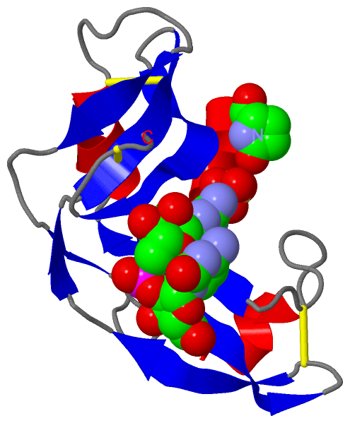
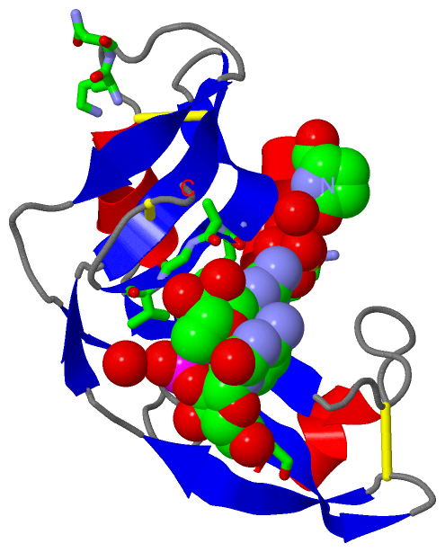
Asymmetric/Biological UnitChain A from PDB Type:PROTEIN Length:105 aligned with Q9DFY5_LITCT | Q9DFY5 from UniProtKB/TrEMBL Length:128 Alignment length:105 33 43 53 63 73 83 93 103 113 123 Q9DFY5_LITCT 24 QDWDTFQKKHLTDTKKVKCDVEMKKALFDCKKTNTFIFARPPRVQALCKNIKDNTNVLSRDVFYLPQCNRKKLPCHYRLDGSTNTICLTCMKELPIHFAGVGKCP 128 SCOP domains d1oj1a_ A: Amphibian cytotoxic ribonuclease SCOP domains CATH domains -1oj1A00 A:2-105 P-30 Protein CATH domains Pfam domains --------------------------------------------------------------------------------------------------------- Pfam domains SAPs(SNPs) --------------------------------------------------------------------------------------------------------- SAPs(SNPs) PROSITE --------------------------------------------------------------------------------------------------------- PROSITE Transcript --------------------------------------------------------------------------------------------------------- Transcript 1oj1 A 1 xDWDTFQKKHLTDTKKVKCDVEMKKALFDCKKTNTFIFARPPRVQALCKNIKNNTNVLSRDVFYLPQCNRKKLPCHYRLDGSTNTICLTCMKELPIHFAGVGKCP 105 | 10 20 30 40 50 60 70 80 90 100 | 1-PCA
|
||||||||||||||||||||
SCOP Domains (1, 1)
Asymmetric/Biological Unit
 |
CATH Domains (1, 1)
Asymmetric/Biological Unit
|
Pfam Domains (0, 0)| (no "Pfam Domain" information available for 1OJ1) |
Gene Ontology (5, 5)|
Asymmetric/Biological Unit(hide GO term definitions) Chain A (Q9DFY5_LITCT | Q9DFY5)
|
||||||||||||||||||||||||||||||||||||||||||
Interactive Views
|
||||||||||||||||||||||||||||||||||||||||||||||||||||||||||||||||||||||||||||||||||||||||||||||||||||||||||||||||||||||||||||||||||||||||||||
Still Images
|
||||||||||||||||
Databases
|
||||||||||||||||||||||||||||||||||||||||||||||||||||||||||||||||||||||||||||||||||||||||||||||||||||||||||||||||||||||||||||||||||||||||||||||||||||||||||||||||
Analysis Tools
|
|||||||||||||||||||||||||||||||||||||||||||||||||||||||||||||
Entries Sharing at Least One Protein Chain (UniProt ID)
Related Entries Specified in the PDB File
|
|