Show PDB file:   
         Plain Text   HTML   (compressed file size)
QuickSearch:   
by PDB,NDB,UniProt,PROSITE Code or Search Term(s)  
(-)Asym./Biol. Unit
(-)Asym./Biol. Unit - sites
collapse expand < >
Image Asym./Biol. Unit
Asym./Biol. Unit  (Jmol Viewer)
Image Asym./Biol. Unit - sites
Asym./Biol. Unit - sites  (Jmol Viewer)

(-) Description

Title :  CRYSTAL STRUCTURE OF CLOSTRIDIUM HISTOLYTICUM COLG COLLAGENASE COLLAGEN-BINDING DOMAIN 3B AT 1.65 ANGSTROM RESOLUTION IN PRESENCE OF CALCIUM
 
Authors :  J. J. Wilson, O. Matsushita, A. Okabe, J. Sakon
Date :  21 Jan 03  (Deposition) - 15 Apr 03  (Release) - 24 Feb 09  (Revision)
Method :  X-RAY DIFFRACTION
Resolution :  1.65
Chains :  Asym./Biol. Unit :  A,B
Keywords :  Beta Sandwich, Metalloprotease, Collagen-Binding Domain, Calcium, Hydrolase (Keyword Search: [Gene Ontology, PubMed, Web (Google))
 
Reference :  J. J. Wilson, O. Matsushita, A. Okabe, J. Sakon
A Bacterial Collagen-Binding Domain With Novel Calcium-Binding Motif Controls Domain Orientation
Embo J. V. 22 1743 2003
PubMed-ID: 12682007  |  Reference-DOI: 10.1093/EMBOJ/CDG172
(for further references see the PDB file header)

(-) Compounds

Molecule 1 - CLASS 1 COLLAGENASE
    ChainsA, B
    EC Number3.4.24.3
    EngineeredYES
    Expression SystemESCHERICHIA COLI BL21
    Expression System PlasmidPGEX-4T-2
    Expression System StrainBL21
    Expression System Taxid511693
    Expression System Vector TypePLASMID
    FragmentCOLLAGEN-BINDING DOMAIN
    GeneCOLG
    Organism ScientificCLOSTRIDIUM HISTOLYTICUM
    Organism Taxid1498

 Structural Features

(-) Chains, Units

  12
Asymmetric/Biological Unit AB

Summary Information (see also Sequences/Alignments below)

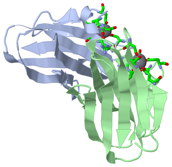
(-) Ligands, Modified Residues, Ions  (1, 4)

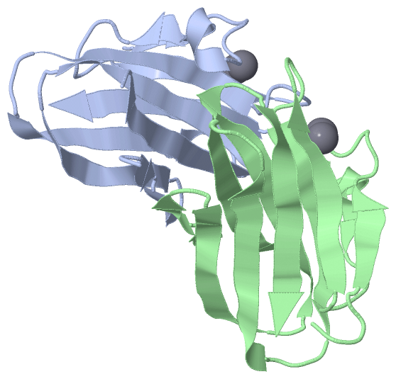
Asymmetric/Biological Unit (1, 4)
No.NameCountTypeFull Name
1CA4Ligand/IonCALCIUM ION

(-) Sites  (4, 4)

Asymmetric Unit (4, 4)
No.NameEvidenceResiduesDescription
1AC1SOFTWAREGLU A:899 , GLU A:901 , SER A:922 , ASP A:927 , ASP A:930 , HOH A:2066BINDING SITE FOR RESIDUE CA A 1009
2AC2SOFTWAREGLU A:901 , ASN A:903 , ASP A:904 , ASP A:927 , ARG A:929 , ASP A:930BINDING SITE FOR RESIDUE CA A 1010
3AC3SOFTWAREGLU B:899 , GLU B:901 , SER B:922 , ASP B:927 , ASP B:930 , HOH B:2126BINDING SITE FOR RESIDUE CA B 1009
4AC4SOFTWAREGLU B:901 , ASN B:903 , ASP B:904 , ASP B:927 , ARG B:929 , ASP B:930BINDING SITE FOR RESIDUE CA B 1010

(-) SS Bonds  (0, 0)

(no "SS Bond" information available for 1NQD)

(-) Cis Peptide Bonds  (2, 2)

Asymmetric/Biological Unit
No.Residues
1Glu A:901 -Asn A:902
2Glu B:901 -Asn B:902

 Sequence-Structure Mapping

(-) SAPs(SNPs)/Variants  (0, 0)

(no "SAP(SNP)/Variant" information available for 1NQD)

(-) PROSITE Motifs  (0, 0)

(no "PROSITE Motif" information available for 1NQD)

(-) Exons   (0, 0)

(no "Exon" information available for 1NQD)

(-) Sequences/Alignments

Asymmetric/Biological Unit
   Reformat: Number of residues per line =  ('0' or empty: single-line sequence representation)
  Number of residues per labelling interval =   
  UniProt sequence: complete  aligned part    
   Show mapping: SCOP domains CATH domains Pfam domains Secondary structure (by author)
SAPs(SNPs) PROSITE motifs Exons
(details for a mapped element are shown in a popup box when the mouse pointer rests over it)
Chain A from PDB  Type:PROTEIN  Length:111
 aligned with Q9S0X0_HATHI | Q9S0X0 from UniProtKB/TrEMBL  Length:1118

    Alignment length:114
                                  1014      1024      1034      1044      1054      1064      1074      1084      1094      1104      1114    
        Q9S0X0_HATHI   1005 EKLKEKENNDSSDKATVIPNFNTTMQGSLLGDDSRDYYSFEVKEEGEVNIELDKKDEFGVTWTLHPESNINDRITYGQVDGNKVSNKVKLRPGKYYLLVYKYSGSGNYELRVNK 1118
               SCOP domains d1nqda_ A: Class 1 collagenase                                                                                     SCOP domains
               CATH domains 1nqdA00 A:895-1008  [code=2.60.120.380, no name defined]                                                           CATH domains
               Pfam domains ------------------------------------------------------------------------------------------------------------------ Pfam domains
         Sec.struct. author .eee......hhhhhee......eeeeee.....eeeeeeee...eeeeeeeee....eeeeeeee..---...ee.eee..eeeeeeee..eeeeeeeeeee..eeeeeeee. Sec.struct. author
                 SAPs(SNPs) ------------------------------------------------------------------------------------------------------------------ SAPs(SNPs)
                    PROSITE ------------------------------------------------------------------------------------------------------------------ PROSITE
                 Transcript ------------------------------------------------------------------------------------------------------------------ Transcript
                1nqd A  895 EKLKEKENNDSSDKATVIPNFNTTMQGSLLGDDSRDYYSFEVKEEGEVNIELDKKDEFGVTWTLHPES---DRITYGQVDGNKVSNKVKLRPGKYYLLVYKYSGSGNYELRVNK 1008
                                   904       914       924       934       944       954       | - |     974       984       994      1004    
                                                                                             962 966                                          

Chain B from PDB  Type:PROTEIN  Length:114
 aligned with Q9S0X0_HATHI | Q9S0X0 from UniProtKB/TrEMBL  Length:1118

    Alignment length:120
                                  1008      1018      1028      1038      1048      1058      1068      1078      1088      1098      1108      1118
        Q9S0X0_HATHI    999 IKGLGNEKLKEKENNDSSDKATVIPNFNTTMQGSLLGDDSRDYYSFEVKEEGEVNIELDKKDEFGVTWTLHPESNINDRITYGQVDGNKVSNKVKLRPGKYYLLVYKYSGSGNYELRVNK 1118
               SCOP domains d1  nqdb_ B: Class 1 collagenase                                                                                         SCOP domains
               CATH domains 1n  qdB00 B:891-1008  [code=2.60.120.380, no name defined]                                                               CATH domains
               Pfam domains ------------------------------------------------------------------------------------------------------------------------ Pfam domains
         Sec.struct. author ..--..eeee......hhhhhee.....eeeeeee.....eeeeeeee...eeeeeeeee....eeeeeeee..----..ee.eee..eeeeeeee..eeeeeeeeeee..eeeeeeee. Sec.struct. author
                 SAPs(SNPs) ------------------------------------------------------------------------------------------------------------------------ SAPs(SNPs)
                    PROSITE ------------------------------------------------------------------------------------------------------------------------ PROSITE
                 Transcript ------------------------------------------------------------------------------------------------------------------------ Transcript
                1nqd B  891 IP--GNEKLKEKENNDSSDKATVIPNFNTTMQGSLLGDDSRDYYSFEVKEEGEVNIELDKKDEFGVTWTLHPES----RITYGQVDGNKVSNKVKLRPGKYYLLVYKYSGSGNYELRVNK 1008
                             |  |  898       908       918       928       938       948       958   |   968       978       988       998      1008
                           892  |                                                                  962  967                                         
                              893                                                                                                                   

   Legend:   → Mismatch (orange background)
  - → Gap (green background, '-', border residues have a numbering label)
    → Modified Residue (blue background, lower-case, 'x' indicates undefined single-letter code, labelled with number + name)
  x → Chemical Group (purple background, 'x', labelled with number + name, e.g. ACE or NH2)
  extra numbering lines below/above indicate numbering irregularities and modified residue names etc., number ends below/above '|'

 Classification and Annotation

(-) SCOP Domains  (1, 2)

Asymmetric/Biological Unit

(-) CATH Domains  (1, 2)

Asymmetric/Biological Unit
(-)
Class: Mainly Beta (13760)

(-) Pfam Domains  (0, 0)

(no "Pfam Domain" information available for 1NQD)

(-) Gene Ontology  (5, 5)

Asymmetric/Biological Unit(hide GO term definitions)
Chain A,B   (Q9S0X0_HATHI | Q9S0X0)
molecular function
    GO:0046872    metal ion binding    Interacting selectively and non-covalently with any metal ion.
    GO:0004252    serine-type endopeptidase activity    Catalysis of the hydrolysis of internal, alpha-peptide bonds in a polypeptide chain by a catalytic mechanism that involves a catalytic triad consisting of a serine nucleophile that is activated by a proton relay involving an acidic residue (e.g. aspartate or glutamate) and a basic residue (usually histidine).
    GO:0008270    zinc ion binding    Interacting selectively and non-covalently with zinc (Zn) ions.
biological process
    GO:0006508    proteolysis    The hydrolysis of proteins into smaller polypeptides and/or amino acids by cleavage of their peptide bonds.
cellular component
    GO:0005576    extracellular region    The space external to the outermost structure of a cell. For cells without external protective or external encapsulating structures this refers to space outside of the plasma membrane. This term covers the host cell environment outside an intracellular parasite.

 Visualization

(-) Interactive Views

Asymmetric/Biological Unit
  Complete Structure
    Jena3D(integrated viewing of ligand, site, SAP, PROSITE, SCOP information)
    WebMol | AstexViewer[tm]@PDBe
(Java Applets, require no local installation except for Java; loading may be slow)
    STRAP
(Java WebStart application, automatic local installation, requires Java; full application with system access!)
    RasMol
(require local installation)
    Molscript (VRML)
(requires installation of a VRML viewer; select preferred view via VRML and generate a mono or stereo PDF format file)
 
  Ligands, Modified Residues, Ions
    CA  [ RasMol | Jena3D ]  +environment [ RasMol | Jena3D ]
 
  Sites
    AC1  [ RasMol ]  +environment [ RasMol ]
    AC2  [ RasMol ]  +environment [ RasMol ]
    AC3  [ RasMol ]  +environment [ RasMol ]
    AC4  [ RasMol ]  +environment [ RasMol ]
 
  Cis Peptide Bonds
    Glu A:901 - Asn A:902   [ RasMol ]  
    Glu B:901 - Asn B:902   [ RasMol ]  
 

(-) Still Images

Jmol
  protein: cartoon or spacefill or dots and stick; nucleic acid: cartoon and stick; ligands: spacefill; active site: stick
Molscript
  protein, nucleic acid: cartoon; ligands: spacefill; active site: ball and stick

 Databases and Analysis Tools

(-) Databases

Access by PDB/NDB ID
  1nqd
    Family and Domain InformationProDom | SYSTERS
    General Structural InformationGlycoscienceDB | MMDB | NDB | OCA | PDB | PDBe | PDBj | PDBsum | PDBWiki | PQS | PROTEOPEDIA
    Orientation in MembranesOPM
    Protein SurfaceSURFACE
    Secondary StructureDSSP (structure derived) | HSSP (homology derived)
    Structural GenomicsGeneCensus
    Structural NeighboursCE | VAST
    Structure ClassificationCATH | Dali | SCOP
    Validation and Original DataBMRB Data View | BMRB Restraints Grid | EDS | PROCHECK | RECOORD | WHAT_CHECK
 
Access by UniProt ID/Accession number
  Q9S0X0_HATHI | Q9S0X0
    Comparative Protein Structure ModelsModBase
    Genomic InformationEnsembl
    Protein-protein InteractionDIP
    Sequence, Family and Domain InformationInterPro | Pfam | SMART | UniProtKB/TrEMBL
 
Access by Enzyme Classificator   (EC Number)
  3.4.24.3
    General Enzyme InformationBRENDA | EC-PDB | Enzyme | IntEnz
    PathwayKEGG | MetaCyc
 
Access by Disease Identifier   (MIM ID)
  (no 'MIM ID' available)
    Disease InformationOMIM
 
Access by GenAge ID
  (no 'GenAge ID' available)
    Age Related InformationGenAge

(-) Analysis Tools

Access by PDB/NDB ID
    Domain InformationXDom
    Interatomic Contacts of Structural UnitsCSU
    Ligand-protein ContactsLPC
    Protein CavitiescastP
    Sequence and Secondary StructurePDBCartoon
    Structure AlignmentSTRAP(Java WebStart application, automatic local installation, requires Java; full application with system access!)
    Structure and Sequence BrowserSTING
 
Access by UniProt ID/Accession number
  Q9S0X0_HATHI | Q9S0X0
    Protein Disorder PredictionDisEMBL | FoldIndex | GLOBPLOT (for more information see DisProt)

 Related Entries

(-) Entries Sharing at Least One Protein Chain (UniProt ID)

UniProtKB/TrEMBL
        Q9S0X0_HATHI | Q9S0X01nqj 2o8o

(-) Related Entries Specified in the PDB File

1nqj