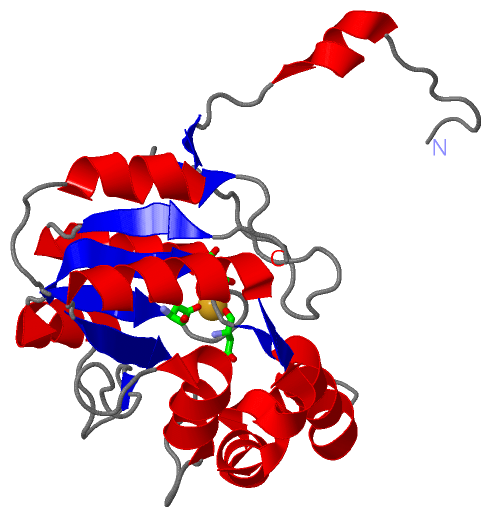
Asymmetric Unit(hide GO term definitions)
Chain A ( APHA_ECOLI | P0AE22)
| molecular function |
|---|
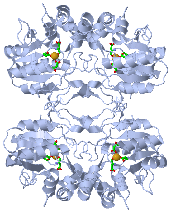
| | GO:0003993 | | acid phosphatase activity | | Catalysis of the reaction: an orthophosphoric monoester + H2O = an alcohol + phosphate, with an acid pH optimum. |
| | GO:0048037 | | cofactor binding | | Interacting selectively and non-covalently with a cofactor, a substance that is required for the activity of an enzyme or other protein. Cofactors may be inorganic, such as the metal atoms zinc, iron, and copper in certain forms, or organic, in which case they are referred to as coenzymes. Cofactors may either be bound tightly to active sites or bind loosely with the substrate. |
| | GO:0016787 | | hydrolase activity | | Catalysis of the hydrolysis of various bonds, e.g. C-O, C-N, C-C, phosphoric anhydride bonds, etc. Hydrolase is the systematic name for any enzyme of EC class 3. |
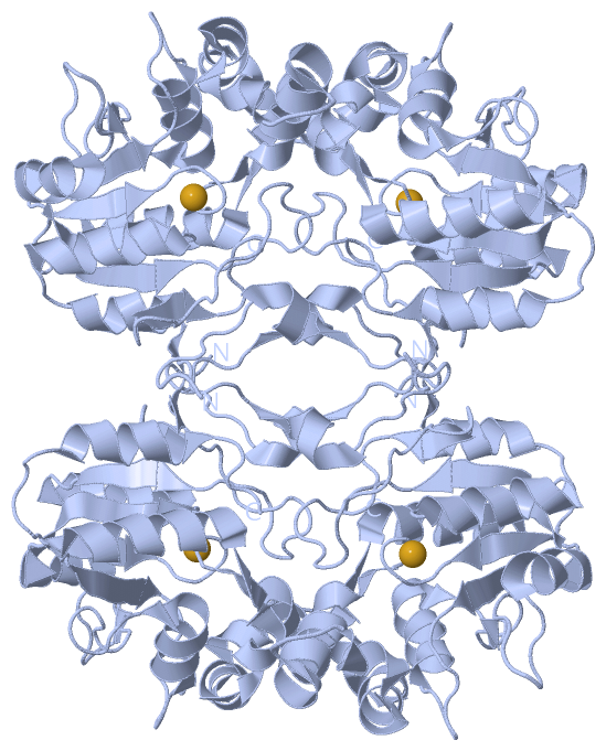
| | GO:0046872 | | metal ion binding | | Interacting selectively and non-covalently with any metal ion. |
| | GO:0004647 | | phosphoserine phosphatase activity | | Catalysis of the reaction: L(or D)-O-phosphoserine + H2O = L(or D)-serine + phosphate. |
| biological process |
|---|
| | GO:0016311 | | dephosphorylation | | The process of removing one or more phosphoric (ester or anhydride) residues from a molecule. |
| cellular component |
|---|
| | GO:0030288 | | outer membrane-bounded periplasmic space | | The region between the inner (cytoplasmic or plasma) membrane and outer membrane of organisms with two membranes such as Gram negative bacteria. These periplasmic spaces are relatively thick and contain a thin peptidoglycan layer (PGL), also referred to as a thin cell wall. |
| | GO:0042597 | | periplasmic space | | The region between the inner (cytoplasmic) and outer membrane (Gram-negative Bacteria) or cytoplasmic membrane and cell wall (Fungi and Gram-positive Bacteria). |
Chain A ( APHA_SHIFL | P0AE23)
| molecular function |
|---|
| | GO:0003993 | | acid phosphatase activity | | Catalysis of the reaction: an orthophosphoric monoester + H2O = an alcohol + phosphate, with an acid pH optimum. |
| | GO:0016787 | | hydrolase activity | | Catalysis of the hydrolysis of various bonds, e.g. C-O, C-N, C-C, phosphoric anhydride bonds, etc. Hydrolase is the systematic name for any enzyme of EC class 3. |
| | GO:0046872 | | metal ion binding | | Interacting selectively and non-covalently with any metal ion. |
| biological process |
|---|
| | GO:0016311 | | dephosphorylation | | The process of removing one or more phosphoric (ester or anhydride) residues from a molecule. |
| cellular component |
|---|
| | GO:0030288 | | outer membrane-bounded periplasmic space | | The region between the inner (cytoplasmic or plasma) membrane and outer membrane of organisms with two membranes such as Gram negative bacteria. These periplasmic spaces are relatively thick and contain a thin peptidoglycan layer (PGL), also referred to as a thin cell wall. |
| | GO:0042597 | | periplasmic space | | The region between the inner (cytoplasmic) and outer membrane (Gram-negative Bacteria) or cytoplasmic membrane and cell wall (Fungi and Gram-positive Bacteria). |
|
 Description