Show PDB file:   
         Plain Text   HTML   (compressed file size)
QuickSearch:   
by PDB,NDB,UniProt,PROSITE Code or Search Term(s)  
(-)Asymmetric Unit
(-)Asym. Unit - sites
(-)Biological Unit 1
(-)Biol. Unit 1 - sites
(-)Biological Unit 2
collapse expand < >
Image Asymmetric Unit
Asymmetric Unit  (Jmol Viewer)
Image Asym. Unit - sites
Asym. Unit - sites  (Jmol Viewer)
Image Biological Unit 1
Biological Unit 1  (Jmol Viewer)
Image Biol. Unit 1 - sites
Biol. Unit 1 - sites  (Jmol Viewer)
Image Biological Unit 2
Biological Unit 2  (Jmol Viewer)

(-) Description

Title :  CRYSTAL STRUCTURE OF CORAL PIGMENT
 
Authors :  M. Prescott, M. Ling, T. Beddoe, A. J. Oakley, S. Dove, O. Hoegh-Guldber R. J. Devenish, J. Rossjohn
Date :  10 Sep 02  (Deposition) - 08 Apr 03  (Release) - 13 Jul 11  (Revision)
Method :  X-RAY DIFFRACTION
Resolution :  2.20
Chains :  Asym. Unit :  A
Biol. Unit 1:  A  (4x)
Biol. Unit 2:  A  (4x)
Keywords :  Blue Coral Pigment, Chromophore, Beta Can Fold, Similar To Green Fluorescent Protein And Dsred, Luminescent Protein (Keyword Search: [Gene Ontology, PubMed, Web (Google))
 
Reference :  M. Prescott, M. Ling, T. Beddoe, A. J. Oakley, S. Dove, O. Hoegh-Guldberg, R. J. Devenish, J. Rossjohn
The 2. 2 A Crystal Structure Of A Pocilloporin Pigment Reveals A Nonplanar Chromophore Conformation.
Structure V. 11 275 2003
PubMed-ID: 12623015  |  Reference-DOI: 10.1016/S0969-2126(03)00028-5

(-) Compounds

Molecule 1 - GFP-LIKE NON-FLUORESCENT CHROMOPROTEIN
    ChainsA
    EngineeredYES
    Expression SystemESCHERICHIA COLI
    Expression System Taxid562
    FragmentRESIDUES 5-225
    Organism ScientificMONTIPORA EFFLORESCENS
    Organism Taxid105610
    SynonymRTMS 5, NON-FLUORESCENT POCILLOPORIN

 Structural Features

(-) Chains, Units

  1
Asymmetric Unit A
Biological Unit 1 (4x)A
Biological Unit 2 (4x)A

Summary Information (see also Sequences/Alignments below)

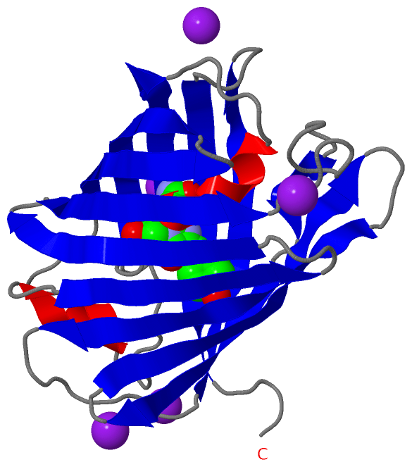
(-) Ligands, Modified Residues, Ions  (2, 6)

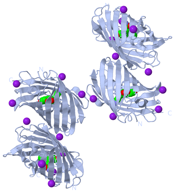
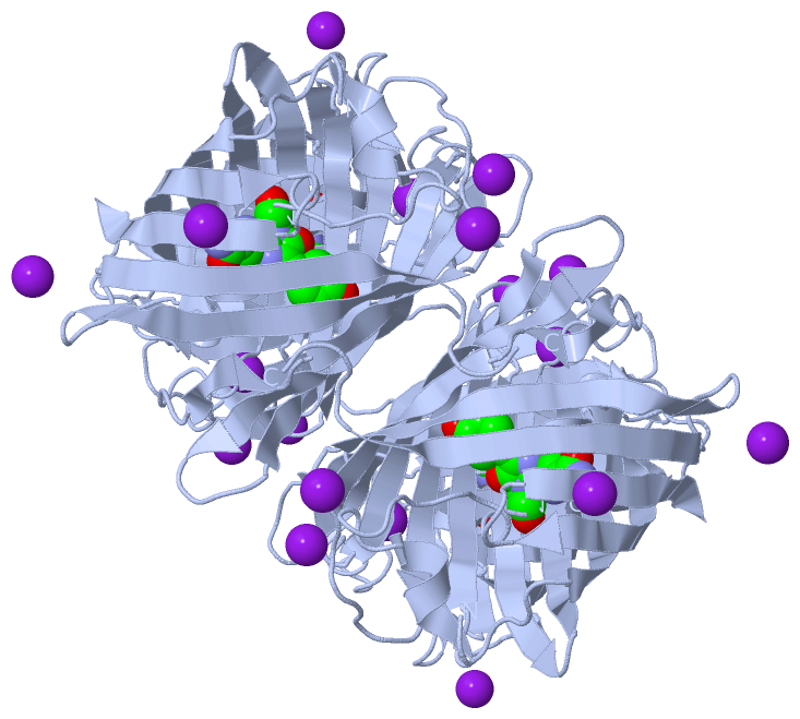
Asymmetric Unit (2, 6)
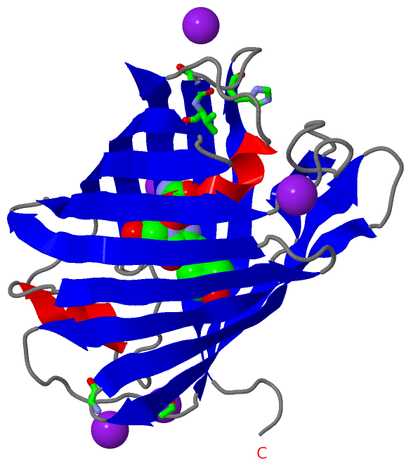
No.NameCountTypeFull Name
1CRQ1Mod. Amino Acid[2-(3-CARBAMOYL-1-IMINO-PROPYL)-4-(4-HYDROXY-BENZYLIDENE)-5-OXO-4,5-DIHYDRO-IMIDAZOL-1-YL]-ACETICACID
2IOD5Ligand/IonIODIDE ION
Biological Unit 1 (2, 24)
No.NameCountTypeFull Name
1CRQ4Mod. Amino Acid[2-(3-CARBAMOYL-1-IMINO-PROPYL)-4-(4-HYDROXY-BENZYLIDENE)-5-OXO-4,5-DIHYDRO-IMIDAZOL-1-YL]-ACETICACID
2IOD20Ligand/IonIODIDE ION
Biological Unit 2 (2, 24)
No.NameCountTypeFull Name
1CRQ4Mod. Amino Acid[2-(3-CARBAMOYL-1-IMINO-PROPYL)-4-(4-HYDROXY-BENZYLIDENE)-5-OXO-4,5-DIHYDRO-IMIDAZOL-1-YL]-ACETICACID
2IOD20Ligand/IonIODIDE ION

(-) Sites  (3, 3)

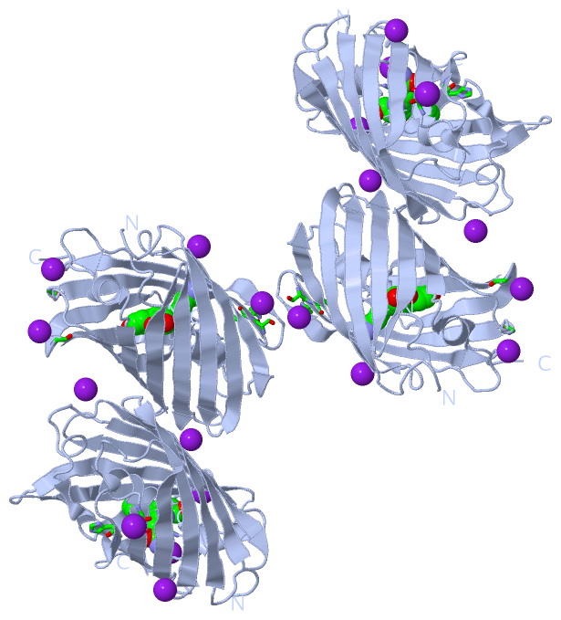
Asymmetric Unit (3, 3)
No.NameEvidenceResiduesDescription
1AC1SOFTWAREHIS A:204 , SER A:210 , VAL A:211BINDING SITE FOR RESIDUE IOD A 301
2AC2SOFTWAREGLY A:155BINDING SITE FOR RESIDUE IOD A 303
3AC3SOFTWAREGLY A:191BINDING SITE FOR RESIDUE IOD A 304

(-) SS Bonds  (0, 0)

(no "SS Bond" information available for 1MOU)

(-) Cis Peptide Bonds  (2, 2)

Asymmetric Unit
No.Residues
1Gly A:52 -Pro A:53
2Phe A:87 -Pro A:88

 Sequence-Structure Mapping

(-) SAPs(SNPs)/Variants  (0, 0)

(no "SAP(SNP)/Variant" information available for 1MOU)

(-) PROSITE Motifs  (0, 0)

(no "PROSITE Motif" information available for 1MOU)

(-) Exons   (0, 0)

(no "Exon" information available for 1MOU)

(-) Sequences/Alignments

Asymmetric Unit
   Reformat: Number of residues per line =  ('0' or empty: single-line sequence representation)
  Number of residues per labelling interval =   
  UniProt sequence: complete  aligned part    
   Show mapping: SCOP domains CATH domains Pfam domains Secondary structure (by author)
SAPs(SNPs) PROSITE motifs Exons
(details for a mapped element are shown in a popup box when the mouse pointer rests over it)
Chain A from PDB  Type:PROTEIN  Length:219
 aligned with NFCP_MONEF | P83690 from UniProtKB/Swiss-Prot  Length:221

    Alignment length:221
                                    10        20        30        40        50        60        70        80        90       100       110       120       130       140       150       160       170       180       190       200       210       220 
           NFCP_MONEF     1 MSVIATQMTYKVYMSGTVNGHYFEVEGDGKGRPYEGEQTVKLTVTKGGPLPFAWDILSPQCQYGSIPFTKYPEDIPDYVKQSFPEGFTWERIMNFEDGAVCTVSNDSSIQGNCFTYHVKFSGLNFPPNGPVMQKKTQGWEPHSERLFARGGMLIGNNFMALKLEGGGHYLCEFKTTYKAKKPVKMPGYHYVDRKLDVTNHNKDYTSVEQCEISIARKPVVA 221
               SCOP domains d1moua_ A: Pocilloporin pigment Rtms5                                                                                                                                                                                         SCOP domains
               CATH domains 1mouA00 A:5-225 Green Fluorescent Protein                                                                                                                                                                                     CATH domains
               Pfam domains --GFP-1mouA01 A:7-222                                                                                                                                                                                                     --- Pfam domains
         Sec.struct. author .......eeeeeeeeeee..eeeeeeeeeeee....eeeeeeeeee......hhhhhhhhh.--............hhhhhhh...eeeeeeeee....eeeeeeeeeee..eeeeeeeeeee............eeee..eeeeeeee..eeeeeeeeeeee....eeeeeeeeeeee.........eeeeeeeeeeee.....eeeeeeeeeee..... Sec.struct. author
                 SAPs(SNPs) ----------------------------------------------------------------------------------------------------------------------------------------------------------------------------------------------------------------------------- SAPs(SNPs)
                    PROSITE ----------------------------------------------------------------------------------------------------------------------------------------------------------------------------------------------------------------------------- PROSITE
                 Transcript ----------------------------------------------------------------------------------------------------------------------------------------------------------------------------------------------------------------------------- Transcript
                 1mou A   5 GGVIATQMTYKVYMSGTVNGHYFEVEGDGKGRPYEGEQTVKLTVTKGGPLPFAWDILSPQCq--SIPFTKYPEDIPDYVKQSFPEGFTWERIMNFEDGAVCTVSNDSSIQGNCFTYHVKFSGLNFPPNGPVMQKKTQGWEPHSERLFARGGMLIGNNFMALKLEGGGHYLCEFKTTYKAKKPVKMPGYHYVDRKLDVTNHNKDYTSVEQCEISIARKPVVA 225
                                    14        24        34        44        54        64 |  |   74        84        94       104       114       124       134       144       154       164       174       184       194       204       214       224 
                                                                                        66-CRQ                                                                                                                                                           

   Legend:   → Mismatch (orange background)
  - → Gap (green background, '-', border residues have a numbering label)
    → Modified Residue (blue background, lower-case, 'x' indicates undefined single-letter code, labelled with number + name)
  x → Chemical Group (purple background, 'x', labelled with number + name, e.g. ACE or NH2)
  extra numbering lines below/above indicate numbering irregularities and modified residue names etc., number ends below/above '|'

 Classification and Annotation

(-) SCOP Domains  (1, 1)

Asymmetric Unit

(-) CATH Domains  (1, 1)

Asymmetric Unit
(-)
Class: Mainly Beta (13760)

(-) Pfam Domains  (1, 1)

Asymmetric Unit
(-)
Clan: GFP (101)

(-) Gene Ontology  (5, 5)

Asymmetric Unit(hide GO term definitions)
Chain A   (NFCP_MONEF | P83690)
molecular function
    GO:0003674    molecular_function    Elemental activities, such as catalysis or binding, describing the actions of a gene product at the molecular level. A given gene product may exhibit one or more molecular functions.
biological process
    GO:0008218    bioluminescence    The production of light by certain enzyme-catalyzed reactions in cells.
    GO:0010117    photoprotection    Protection mechanism used by plants under conditions of excess energy absorption as a consequence of the light reactions of photosynthesis.
    GO:0018298    protein-chromophore linkage    The covalent or noncovalent attachment of a chromophore to a protein.
cellular component
    GO:0018995    host    Any organism in which another organism, especially a parasite or symbiont, spends part or all of its life cycle and from which it obtains nourishment and/or protection.

 Visualization

(-) Interactive Views

Asymmetric Unit
  Complete Structure
    Jena3D(integrated viewing of ligand, site, SAP, PROSITE, SCOP information)
    WebMol | AstexViewer[tm]@PDBe
(Java Applets, require no local installation except for Java; loading may be slow)
    STRAP
(Java WebStart application, automatic local installation, requires Java; full application with system access!)
    RasMol
(require local installation)
    Molscript (VRML)
(requires installation of a VRML viewer; select preferred view via VRML and generate a mono or stereo PDF format file)
 
  Ligands, Modified Residues, Ions
    CRQ  [ RasMol | Jena3D ]  +environment [ RasMol | Jena3D ]
    IOD  [ RasMol | Jena3D ]  +environment [ RasMol | Jena3D ]
 
  Sites
    AC1  [ RasMol ]  +environment [ RasMol ]
    AC2  [ RasMol ]  +environment [ RasMol ]
    AC3  [ RasMol ]  +environment [ RasMol ]
 
  Cis Peptide Bonds
    Gly A:52 - Pro A:53   [ RasMol ]  
    Phe A:87 - Pro A:88   [ RasMol ]  
 
Biological Units
  Complete Structure
    Biological Unit 1  [ Jena3D ]
    Biological Unit 2  [ Jena3D ]

(-) Still Images

Jmol
  protein: cartoon or spacefill or dots and stick; nucleic acid: cartoon and stick; ligands: spacefill; active site: stick
Molscript
  protein, nucleic acid: cartoon; ligands: spacefill; active site: ball and stick

 Databases and Analysis Tools

(-) Databases

Access by PDB/NDB ID
  1mou
    Family and Domain InformationProDom | SYSTERS
    General Structural InformationGlycoscienceDB | MMDB | NDB | OCA | PDB | PDBe | PDBj | PDBsum | PDBWiki | PQS | PROTEOPEDIA
    Orientation in MembranesOPM
    Protein SurfaceSURFACE
    Secondary StructureDSSP (structure derived) | HSSP (homology derived)
    Structural GenomicsGeneCensus
    Structural NeighboursCE | VAST
    Structure ClassificationCATH | Dali | SCOP
    Validation and Original DataBMRB Data View | BMRB Restraints Grid | EDS | PROCHECK | RECOORD | WHAT_CHECK
 
Access by UniProt ID/Accession number
  NFCP_MONEF | P83690
    Comparative Protein Structure ModelsModBase
    Genomic InformationEnsembl
    Protein-protein InteractionDIP
    Sequence, Family and Domain InformationInterPro | Pfam | SMART | UniProtKB/SwissProt
 
Access by Enzyme Classificator   (EC Number)
  (no 'Enzyme Classificator' available)
    General Enzyme InformationBRENDA | EC-PDB | Enzyme | IntEnz
    PathwayKEGG | MetaCyc
 
Access by Disease Identifier   (MIM ID)
  (no 'MIM ID' available)
    Disease InformationOMIM
 
Access by GenAge ID
  (no 'GenAge ID' available)
    Age Related InformationGenAge

(-) Analysis Tools

Access by PDB/NDB ID
    Domain InformationXDom
    Interatomic Contacts of Structural UnitsCSU
    Ligand-protein ContactsLPC
    Protein CavitiescastP
    Sequence and Secondary StructurePDBCartoon
    Structure AlignmentSTRAP(Java WebStart application, automatic local installation, requires Java; full application with system access!)
    Structure and Sequence BrowserSTING
 
Access by UniProt ID/Accession number
  NFCP_MONEF | P83690
    Protein Disorder PredictionDisEMBL | FoldIndex | GLOBPLOT (for more information see DisProt)

 Related Entries

(-) Entries Sharing at Least One Protein Chain (UniProt ID)

UniProtKB/Swiss-Prot
        NFCP_MONEF | P836901mov 2arl 2p4m 3vic 3vk1

(-) Related Entries Specified in the PDB File

1mov 1MOV CONTAINS CRYSTAL STRUCTURE OF THE SAME PROTEIN COMPLEXED WITH H146S