| molecular function |
|---|
| | GO:0005070 | | SH3/SH2 adaptor activity | | Interacting selectively and non-covalently and simultaneously with one or more signal transduction molecules, usually acting as a scaffold to bring these molecules into close proximity either using their own SH2/SH3 domains (e.g. Grb2) or those of their target molecules (e.g. SAM68). |
| | GO:0005515 | | protein binding | | Interacting selectively and non-covalently with any protein or protein complex (a complex of two or more proteins that may include other nonprotein molecules). |
| biological process |
|---|
| | GO:0042113 | | B cell activation | | The change in morphology and behavior of a mature or immature B cell resulting from exposure to a mitogen, cytokine, chemokine, cellular ligand, or an antigen for which it is specific. |
| | GO:0002757 | | immune response-activating signal transduction | | The cascade of processes by which a signal interacts with a receptor, causing a change in the level or activity of a second messenger or other downstream target, and ultimately leading to activation or perpetuation of an immune response. |
| | GO:0008285 | | negative regulation of cell proliferation | | Any process that stops, prevents or reduces the rate or extent of cell proliferation. |
| | GO:0009967 | | positive regulation of signal transduction | | Any process that activates or increases the frequency, rate or extent of signal transduction. |
| cellular component |
|---|
| | GO:0005737 | | cytoplasm | | All of the contents of a cell excluding the plasma membrane and nucleus, but including other subcellular structures. |
| | GO:0005654 | | nucleoplasm | | That part of the nuclear content other than the chromosomes or the nucleolus. |
| | GO:0005886 | | plasma membrane | | The membrane surrounding a cell that separates the cell from its external environment. It consists of a phospholipid bilayer and associated proteins. |
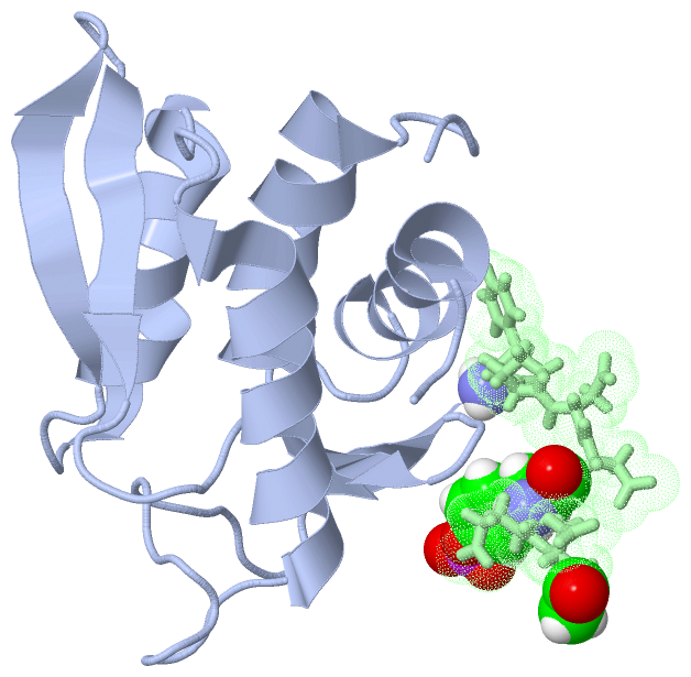
 Description
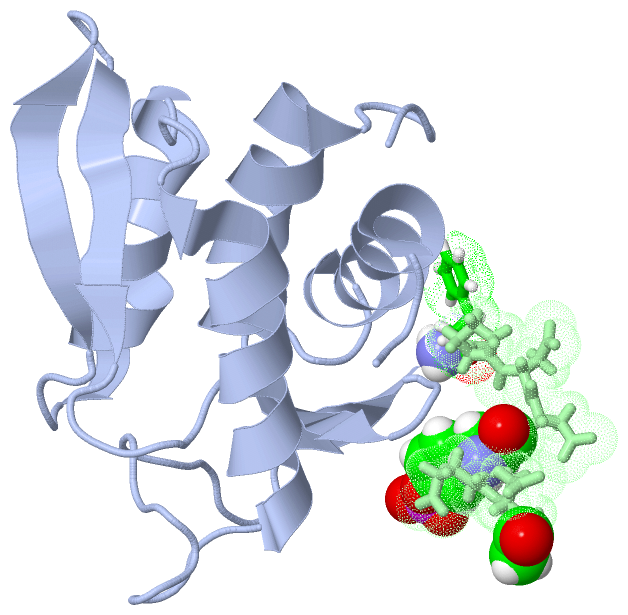