Show PDB file:   
         Plain Text   HTML   (compressed file size)
QuickSearch:   
by PDB,NDB,UniProt,PROSITE Code or Search Term(s)  
(-)Asym./Biol. Unit
(-)Asym./Biol. Unit - sites
collapse expand < >
Image Asym./Biol. Unit
Asym./Biol. Unit  (Jmol Viewer)
Image Asym./Biol. Unit - sites
Asym./Biol. Unit - sites  (Jmol Viewer)

(-) Description

Title :  CRYSTAL STRUCTURE OF CHROMOBACTERIUM VIOLACEUM PHENYLALANINE HYDROXYLASE, STRUCTURE HAS BOUND IRON (III) AND OXIDIZED COFACTOR 7,8-DIHYDROBIOPTERIN
 
Authors :  H. Erlandsen, J. Y. Kim, M. G. Patch, A. Han, A. Volner, M. M. Abu-Omar, R. C. Stevens
Date :  21 May 02  (Deposition) - 17 Jul 02  (Release) - 24 Feb 09  (Revision)
Method :  X-RAY DIFFRACTION
Resolution :  1.40
Chains :  Asym./Biol. Unit :  A
Keywords :  Oxidoreductase, Hydroxylase, Phenylalanine, Dihydrobiopterin, Bacterial Enzyme (Keyword Search: [Gene Ontology, PubMed, Web (Google))
 
Reference :  H. Erlandsen, J. Y. Kim, M. G. Patch, A. Han, A. Volner, M. M. Abu-Omar, R. C. Stevens
Structural Comparison Of Bacterial And Human Iron-Dependent Phenylalanine Hydroxylases: Similar Fold, Different Stability And Reaction Rates.
J. Mol. Biol. V. 320 645 2002
PubMed-ID: 12096915  |  Reference-DOI: 10.1016/S0022-2836(02)00496-5
(for further references see the PDB file header)

(-) Compounds

Molecule 1 - PHENYLALANINE-4-HYDROXYLASE
    ChainsA
    EC Number1.14.16.1
    EngineeredYES
    Expression SystemESCHERICHIA COLI
    Expression System PlasmidPET-3A
    Expression System StrainBL21(DE3)PLYSS
    Expression System Taxid562
    Expression System Vector TypePLASMID
    Organism ScientificCHROMOBACTERIUM VIOLACEUM
    Organism Taxid536
    SynonymPAH, PHE-4-MONOOXYGENASE, PHENYLALANINASE

 Structural Features

(-) Chains, Units

  1
Asymmetric/Biological Unit A

Summary Information (see also Sequences/Alignments below)

(-) Ligands, Modified Residues, Ions  (3, 3)

Asymmetric/Biological Unit (3, 3)
No.NameCountTypeFull Name
1CL1Ligand/IonCHLORIDE ION
2FE1Ligand/IonFE (III) ION
3HBI1Ligand/Ion7,8-DIHYDROBIOPTERIN

(-) Sites  (3, 3)

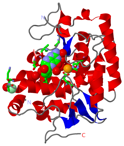
Asymmetric Unit (3, 3)
No.NameEvidenceResiduesDescription
1AC1SOFTWAREHIS A:138 , HIS A:143 , GLU A:184 , HOH A:1002 , HOH A:1003BINDING SITE FOR RESIDUE FE A 400
2AC2SOFTWARELYS A:167 , ALA A:168 , HOH A:1078BINDING SITE FOR RESIDUE CL A 401
3AC3SOFTWAREGLY A:100 , ILE A:102 , ASP A:104 , PHE A:107 , TYR A:159 , HOH A:1001 , HOH A:1002 , HOH A:1004 , HOH A:1005 , HOH A:1006 , HOH A:1030BINDING SITE FOR RESIDUE HBI A 500

(-) SS Bonds  (0, 0)

(no "SS Bond" information available for 1LTZ)

(-) Cis Peptide Bonds  (0, 0)

(no "Cis Peptide Bond" information available for 1LTZ)

 Sequence-Structure Mapping

(-) SAPs(SNPs)/Variants  (0, 0)

(no "SAP(SNP)/Variant" information available for 1LTZ)

(-) PROSITE Motifs  (1, 1)

Asymmetric/Biological Unit (1, 1)
 PROSITEUniProtKBPDB
No.IDACDescriptionIDLocationCountLocation
1BH4_AAA_HYDROXYL_1PS00367 Biopterin-dependent aromatic amino acid hydroxylases signature.PH4H_CHRVO134-145  1A:134-145

(-) Exons   (0, 0)

(no "Exon" information available for 1LTZ)

(-) Sequences/Alignments

Asymmetric/Biological Unit
   Reformat: Number of residues per line =  ('0' or empty: single-line sequence representation)
  Number of residues per labelling interval =   
  UniProt sequence: complete  aligned part    
   Show mapping: SCOP domains CATH domains Pfam domains Secondary structure (by author)
SAPs(SNPs) PROSITE motifs Exons
(details for a mapped element are shown in a popup box when the mouse pointer rests over it)
Chain A from PDB  Type:PROTEIN  Length:274
 aligned with PH4H_CHRVO | P30967 from UniProtKB/Swiss-Prot  Length:297

    Alignment length:277
                                    16        26        36        46        56        66        76        86        96       106       116       126       136       146       156       166       176       186       196       206       216       226       236       246       256       266       276       
           PH4H_CHRVO     7 FVVPDITTRKNVGLSHDANDFTLPQPLDRYSAEDHATWATLYQRQCKLLPGRACDEFMEGLERLEVDADRVPDFNKLNQKLMAATGWKIVAVPGLIPDDVFFEHLANRRFPVTWWLREPHQLDYLQEPDVFHDLFGHVPLLINPVFADYLEAYGKGGVKAKALGALPMLARLYWYTVEFGLINTPAGMRIYGAGILSSKSESIYCLDSASPNRVGFDLMRIMNTRYRIDTFQKTYFVIDSFKQLFDATAPDFAPLYLQLADAQPWGAGDVAPDDLVL 283
               SCOP domains d1ltza_ A: Phenylalanine hydroxylase, PAH                                                                                                                                                                                                                                             SCOP domains
               CATH domains 1ltzA00 A:7-283 Phenylalanine Hydroxylase                                                                                                                                                                                                                                             CATH domains
               Pfam domains -------Biopterin_H-1ltzA01 A:14-259                                                                                                                                                                                                                          ------------------------ Pfam domains
         Sec.struct. author ....hhhhhhh..............hhhhhhhhhhhhhhhhhhhhhhhh....hhhhhhhhhhhh.......hhhhhhhhhhhhhh.eeeee....hhhhhhhhhhh.eeeee...............hhhhhhhhhhhhhhhhhhhhhhhhhhhhhhhh...hhhhhhhhhhhhh....eeee..eeee.hhhhhhhhhhhhhh......eeee.hhhhhhh..........eeeee.hhhhhhhh---.hhhhhhhhh................. Sec.struct. author
                 SAPs(SNPs) ------------------------------------------------------------------------------------------------------------------------------------------------------------------------------------------------------------------------------------------------------------------------------------- SAPs(SNPs)
                    PROSITE -------------------------------------------------------------------------------------------------------------------------------BH4_AAA_HYDR------------------------------------------------------------------------------------------------------------------------------------------ PROSITE
                 Transcript ------------------------------------------------------------------------------------------------------------------------------------------------------------------------------------------------------------------------------------------------------------------------------------- Transcript
                 1ltz A   7 FVVPDITTRKNVGLSHDANDFTLPQPLDRYSAEDHATWATLYQRQCKLLPGRACDEFLEGLERLEVDADRVPDFNKLNEKLMAATGWKIVAVPGLIPDDVFFEHLANRRFPVTWWLREPHQLDYLQEPDVFHDLFGHVPLLINPVFADYLEAYGKGGVKAKALGALPMLARLYWYTVEFGLINTPAGMRIYGAGILSSKSESIYCLDSASPNRVGFDLMRIMNTRYRIDTFQKTYFVIDSFKQLFDA---DFAPLYLQLADAQPWGAGDIAPDDLVL 283
                                    16        26        36        46        56        66        76        86        96       106       116       126       136       146       156       166       176       186       196       206       216       226       236       246      |  -|      266       276       
                                                                                                                                                                                                                                                                                253 257                          

   Legend:   → Mismatch (orange background)
  - → Gap (green background, '-', border residues have a numbering label)
    → Modified Residue (blue background, lower-case, 'x' indicates undefined single-letter code, labelled with number + name)
  x → Chemical Group (purple background, 'x', labelled with number + name, e.g. ACE or NH2)
  extra numbering lines below/above indicate numbering irregularities and modified residue names etc., number ends below/above '|'

 Classification and Annotation

(-) SCOP Domains  (1, 1)

Asymmetric/Biological Unit

(-) CATH Domains  (1, 1)

Asymmetric/Biological Unit

(-) Pfam Domains  (1, 1)

Asymmetric/Biological Unit

(-) Gene Ontology  (9, 9)

Asymmetric/Biological Unit(hide GO term definitions)
Chain A   (PH4H_CHRVO | P30967)
molecular function
    GO:0005506    iron ion binding    Interacting selectively and non-covalently with iron (Fe) ions.
    GO:0046872    metal ion binding    Interacting selectively and non-covalently with any metal ion.
    GO:0004497    monooxygenase activity    Catalysis of the incorporation of one atom from molecular oxygen into a compound and the reduction of the other atom of oxygen to water.
    GO:0016491    oxidoreductase activity    Catalysis of an oxidation-reduction (redox) reaction, a reversible chemical reaction in which the oxidation state of an atom or atoms within a molecule is altered. One substrate acts as a hydrogen or electron donor and becomes oxidized, while the other acts as hydrogen or electron acceptor and becomes reduced.
    GO:0016714    oxidoreductase activity, acting on paired donors, with incorporation or reduction of molecular oxygen, reduced pteridine as one donor, and incorporation of one atom of oxygen    Catalysis of an oxidation-reduction (redox) reaction in which hydrogen or electrons are transferred from reduced pteridine and one other donor, and one atom of oxygen is incorporated into one donor.
    GO:0004505    phenylalanine 4-monooxygenase activity    Catalysis of the reaction: L-phenylalanine + tetrahydrobiopterin + O2 = L-tyrosine + 4-alpha-hydroxytetrahydrobiopterin.
biological process
    GO:0006559    L-phenylalanine catabolic process    The chemical reactions and pathways resulting in the breakdown of phenylalanine, 2-amino-3-phenylpropanoic acid.
    GO:0009072    aromatic amino acid family metabolic process    The chemical reactions and pathways involving aromatic amino acid family, amino acids with aromatic ring (phenylalanine, tyrosine, tryptophan).
    GO:0055114    oxidation-reduction process    A metabolic process that results in the removal or addition of one or more electrons to or from a substance, with or without the concomitant removal or addition of a proton or protons.

 Visualization

(-) Interactive Views

Asymmetric/Biological Unit
  Complete Structure
    Jena3D(integrated viewing of ligand, site, SAP, PROSITE, SCOP information)
    WebMol | AstexViewer[tm]@PDBe
(Java Applets, require no local installation except for Java; loading may be slow)
    STRAP
(Java WebStart application, automatic local installation, requires Java; full application with system access!)
    RasMol
(require local installation)
    Molscript (VRML)
(requires installation of a VRML viewer; select preferred view via VRML and generate a mono or stereo PDF format file)
 
  Ligands, Modified Residues, Ions
    CL  [ RasMol | Jena3D ]  +environment [ RasMol | Jena3D ]
    FE  [ RasMol | Jena3D ]  +environment [ RasMol | Jena3D ]
    HBI  [ RasMol | Jena3D ]  +environment [ RasMol | Jena3D ]
 
  Sites
    AC1  [ RasMol ]  +environment [ RasMol ]
    AC2  [ RasMol ]  +environment [ RasMol ]
    AC3  [ RasMol ]  +environment [ RasMol ]
 
  Cis Peptide Bonds
(no "Cis Peptide Bonds" information available for 1ltz)
 

(-) Still Images

Jmol
  protein: cartoon or spacefill or dots and stick; nucleic acid: cartoon and stick; ligands: spacefill; active site: stick
Molscript
  protein, nucleic acid: cartoon; ligands: spacefill; active site: ball and stick

 Databases and Analysis Tools

(-) Databases

Access by PDB/NDB ID
  1ltz
    Family and Domain InformationProDom | SYSTERS
    General Structural InformationGlycoscienceDB | MMDB | NDB | OCA | PDB | PDBe | PDBj | PDBsum | PDBWiki | PQS | PROTEOPEDIA
    Orientation in MembranesOPM
    Protein SurfaceSURFACE
    Secondary StructureDSSP (structure derived) | HSSP (homology derived)
    Structural GenomicsGeneCensus
    Structural NeighboursCE | VAST
    Structure ClassificationCATH | Dali | SCOP
    Validation and Original DataBMRB Data View | BMRB Restraints Grid | EDS | PROCHECK | RECOORD | WHAT_CHECK
 
Access by UniProt ID/Accession number
  PH4H_CHRVO | P30967
    Comparative Protein Structure ModelsModBase
    Genomic InformationEnsembl
    Protein-protein InteractionDIP
    Sequence, Family and Domain InformationInterPro | Pfam | SMART | UniProtKB/SwissProt
 
Access by Enzyme Classificator   (EC Number)
  1.14.16.1
    General Enzyme InformationBRENDA | EC-PDB | Enzyme | IntEnz
    PathwayKEGG | MetaCyc
 
Access by Disease Identifier   (MIM ID)
  (no 'MIM ID' available)
    Disease InformationOMIM
 
Access by GenAge ID
  (no 'GenAge ID' available)
    Age Related InformationGenAge

(-) Analysis Tools

Access by PDB/NDB ID
    Domain InformationXDom
    Interatomic Contacts of Structural UnitsCSU
    Ligand-protein ContactsLPC
    Protein CavitiescastP
    Sequence and Secondary StructurePDBCartoon
    Structure AlignmentSTRAP(Java WebStart application, automatic local installation, requires Java; full application with system access!)
    Structure and Sequence BrowserSTING
 
Access by UniProt ID/Accession number
  PH4H_CHRVO | P30967
    Protein Disorder PredictionDisEMBL | FoldIndex | GLOBPLOT (for more information see DisProt)

 Related Entries

(-) Entries Sharing at Least One Protein Chain (UniProt ID)

UniProtKB/Swiss-Prot
        PH4H_CHRVO | P309671ltu 1ltv 3tcy 3tk2 3tk4 4esm 4etl 4jpx 4jpy 4q3w 4q3x 4q3y 4q3z

(-) Related Entries Specified in the PDB File

1ltu CRYSTAL STRUCTURE OF CHROMOBACTERIUM VIOLACEUM, APO (NO IRON BOUND) STRUCTURE
1ltv CRYSTAL STRUCTURE CHROMOBACTERIUM VIOLACEUM PHENYLALANINE HYDROXYLASE, STRUCTURE WITH BOUND OXIDIZED FE(III)