Show PDB file:   
         Plain Text   HTML   (compressed file size)
QuickSearch:   
by PDB,NDB,UniProt,PROSITE Code or Search Term(s)  
(-)Asym./Biol. Unit
(-)Asym./Biol. Unit - sites
collapse expand < >
Image Asym./Biol. Unit
Asym./Biol. Unit  (Jmol Viewer)
Image Asym./Biol. Unit - sites
Asym./Biol. Unit - sites  (Jmol Viewer)

(-) Description

Title :  CRYSTAL STRUCTURE OF THE FOSFOMYCIN RESISTANCE PROTEIN (FOSA) CONTAINING BOUND SUBSTRATE
 
Authors :  C. L. Rife, R. E. Pharris, M. E. Newcomer, R. N. Armstrong
Date :  11 May 02  (Deposition) - 11 Sep 02  (Release) - 24 Feb 09  (Revision)
Method :  X-RAY DIFFRACTION
Resolution :  1.19
Chains :  Asym./Biol. Unit :  A,B
Keywords :  Potassium Binding Loop, Manganese Binding, Transferase (Keyword Search: [Gene Ontology, PubMed, Web (Google))
 
Reference :  C. L. Rife, R. E. Pharris, M. E. Newcomer, R. N. Armstrong
Crystal Structure Of A Genomically Encoded Fosfomycin Resistance Protein (Fosa) At 1. 19 A Resolution By Mad Phasing Off The L-Iii Edge Of Tl(+)
J. Am. Chem. Soc. V. 124 11001 2002
PubMed-ID: 12224946  |  Reference-DOI: 10.1021/JA026879V
(for further references see the PDB file header)

(-) Compounds

Molecule 1 - PROBABLE FOSFOMYCIN RESISTANCE PROTEIN
    ChainsA, B
    EC Number2.5.1.18
    EngineeredYES
    Expression SystemESCHERICHIA COLI BL21(DE3)
    Expression System PlasmidPET-20
    Expression System StrainBL21-DE3
    Expression System Taxid469008
    Expression System Vector TypePLASMID
    GeneFOSA
    Organism ScientificPSEUDOMONAS AERUGINOSA
    Organism Taxid287
    SynonymFOSA

 Structural Features

(-) Chains, Units

  12
Asymmetric/Biological Unit AB

Summary Information (see also Sequences/Alignments below)
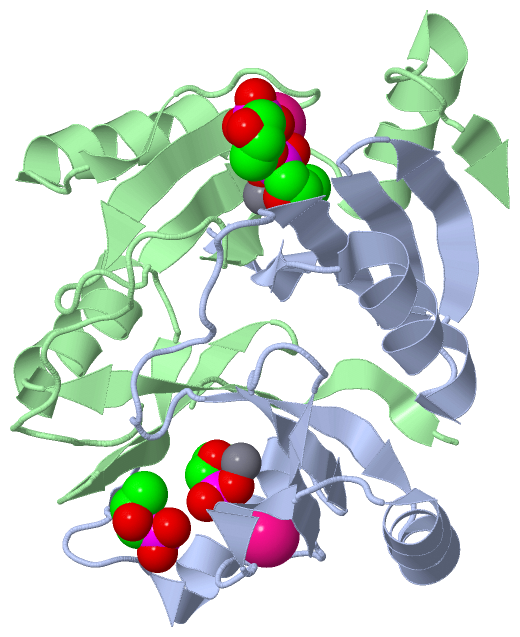
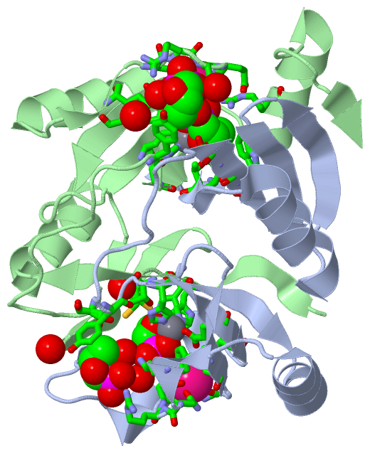
(-) Ligands, Modified Residues, Ions  (3, 8)

Asymmetric/Biological Unit (3, 8)
No.NameCountTypeFull Name
1FCN4Ligand/IonFOSFOMYCIN
2K2Ligand/IonPOTASSIUM ION
3MN2Ligand/IonMANGANESE (II) ION

(-) Sites  (8, 8)

Asymmetric Unit (8, 8)
No.NameEvidenceResiduesDescription
1AC1SOFTWAREASN B:92 , SER B:94 , GLU B:95 , GLY B:96 , SER B:98 , HIS B:112 , ARG B:119 , HOH B:9014BINDING SITE FOR RESIDUE K B 3001
2AC2SOFTWAREASN A:92 , SER A:94 , GLU A:95 , GLY A:96 , SER A:98 , HIS A:112 , ARG A:119 , HOH A:9016BINDING SITE FOR RESIDUE K A 3002
3AC3SOFTWAREHIS A:7 , HIS B:64 , GLU B:110 , FCN B:4001BINDING SITE FOR RESIDUE MN B 3003
4AC4SOFTWAREHIS A:64 , GLU A:110 , FCN A:4002 , HIS B:7BINDING SITE FOR RESIDUE MN A 3004
5AC5SOFTWAREHIS A:7 , THR A:9 , TRP A:46 , CYS A:48 , HIS B:64 , LYS B:90 , SER B:94 , TYR B:100 , GLU B:110 , ARG B:119 , MN B:3003 , HOH B:9014 , HOH B:9078 , HOH B:9095BINDING SITE FOR RESIDUE FCN B 4001
6AC6SOFTWAREHIS A:64 , LYS A:90 , SER A:94 , TYR A:100 , GLU A:110 , ARG A:119 , MN A:3004 , HOH A:9015 , HOH A:9016 , HOH A:9080 , HIS B:7 , THR B:9 , TRP B:46 , CYS B:48BINDING SITE FOR RESIDUE FCN A 4002
7AC7SOFTWARESER A:50 , TYR A:62 , FCN A:4004 , TYR B:62 , LYS B:90 , ARG B:93 , HOH B:9078 , HOH B:9095 , HOH B:9328 , HOH B:9339 , HOH B:9364BINDING SITE FOR RESIDUE FCN B 4003
8AC8SOFTWARETYR A:62 , LYS A:90 , ARG A:93 , HOH A:9015 , HOH A:9080 , HOH A:9223 , HOH A:9246 , SER B:50 , TYR B:62 , FCN B:4003BINDING SITE FOR RESIDUE FCN A 4004

(-) SS Bonds  (0, 0)

(no "SS Bond" information available for 1LQP)

(-) Cis Peptide Bonds  (0, 0)

(no "Cis Peptide Bond" information available for 1LQP)

 Sequence-Structure Mapping

(-) SAPs(SNPs)/Variants  (0, 0)

(no "SAP(SNP)/Variant" information available for 1LQP)

(-) PROSITE Motifs  (1, 2)

Asymmetric/Biological Unit (1, 2)
 PROSITEUniProtKBPDB
No.IDACDescriptionIDLocationCountLocation
1VOCPS51819 Vicinal oxygen chelate (VOC) domain profile.FOSA_PSEAE4-114
 
  2A:4-114
B:4-114

(-) Exons   (0, 0)

(no "Exon" information available for 1LQP)

(-) Sequences/Alignments

Asymmetric/Biological Unit
   Reformat: Number of residues per line =  ('0' or empty: single-line sequence representation)
  Number of residues per labelling interval =   
  UniProt sequence: complete  aligned part    
   Show mapping: SCOP domains CATH domains Pfam domains Secondary structure (by author)
SAPs(SNPs) PROSITE motifs Exons
(details for a mapped element are shown in a popup box when the mouse pointer rests over it)
Chain A from PDB  Type:PROTEIN  Length:134
 aligned with FOSA_PSEAE | Q9I4K6 from UniProtKB/Swiss-Prot  Length:135

    Alignment length:134
                                    10        20        30        40        50        60        70        80        90       100       110       120       130    
           FOSA_PSEAE     1 MLTGLNHLTLAVADLPASIAFYRDLLGFRLEARWDQGAYLELGSLWLCLSREPQYGGPAADYTHYAFGIAAADFARFAAQLRAHGVREWKQNRSEGDSFYFLDPDGHRLEAHVGDLRSRLAACRQAPYAGMRFA 134
               SCOP domains d1lqpa_ A: Fosfomycin resistance protein A (FosA)                                                                                      SCOP domains
               CATH domains 1lqpA00 A:1-134 2,3-Dihydroxybiphenyl 1,2-Dioxygenase, domain 1                                                                        CATH domains
               Pfam domains -------------------------------------------------------------------------------------------------------------------------------------- Pfam domains
         Sec.struct. author .eeeeeeeeeee.hhhhhhhhhhhh...eeeeee..eeeeee..eeeeeee............eeeeee...hhhhhhhhhhhh...eee.......eeeee.....eeeee..hhhhhhhhhhhh.....ee. Sec.struct. author
                 SAPs(SNPs) -------------------------------------------------------------------------------------------------------------------------------------- SAPs(SNPs)
                    PROSITE ---VOC  PDB: A:4-114 UniProt: 4-114                                                                               -------------------- PROSITE
                 Transcript -------------------------------------------------------------------------------------------------------------------------------------- Transcript
                 1lqp A   1 MLTGLNHLTLAVADLPASIAFYRDLLGFRLEARWDQGAYLELGSLWLCLSREPQYGGPAADYTHYAFGIAAADFARFAAQLRAHGVREWKQNRSEGDSFYFLDPDGHRLEAHVGDLRSRLAACRQAPYAGMRFA 134
                                    10        20        30        40        50        60        70        80        90       100       110       120       130    

Chain B from PDB  Type:PROTEIN  Length:134
 aligned with FOSA_PSEAE | Q9I4K6 from UniProtKB/Swiss-Prot  Length:135

    Alignment length:134
                                    10        20        30        40        50        60        70        80        90       100       110       120       130    
           FOSA_PSEAE     1 MLTGLNHLTLAVADLPASIAFYRDLLGFRLEARWDQGAYLELGSLWLCLSREPQYGGPAADYTHYAFGIAAADFARFAAQLRAHGVREWKQNRSEGDSFYFLDPDGHRLEAHVGDLRSRLAACRQAPYAGMRFA 134
               SCOP domains d1lqpb_ B: Fosfomycin resistance protein A (FosA)                                                                                      SCOP domains
               CATH domains 1lqpB00 B:1-134 2,3-Dihydroxybiphenyl 1,2-Dioxygenase, domain 1                                                                        CATH domains
           Pfam domains (1) ---------Glyoxalase_2-1lqpB01 B:10-112                                                                          ---------------------- Pfam domains (1)
           Pfam domains (2) ---------Glyoxalase_2-1lqpB02 B:10-112                                                                          ---------------------- Pfam domains (2)
         Sec.struct. author .eeeeeeeeeee.hhhhhhhhhhhh...eeeeee..eeeeee..eeeeeee............eeeeee...hhhhhhhhhhhh...eee.......eeeee.....eeeee..hhhhhhhhhhhh.....ee. Sec.struct. author
                 SAPs(SNPs) -------------------------------------------------------------------------------------------------------------------------------------- SAPs(SNPs)
                    PROSITE ---VOC  PDB: B:4-114 UniProt: 4-114                                                                               -------------------- PROSITE
                 Transcript -------------------------------------------------------------------------------------------------------------------------------------- Transcript
                 1lqp B   1 MLTGLNHLTLAVADLPASIAFYRDLLGFRLEARWDQGAYLELGSLWLCLSREPQYGGPAADYTHYAFGIAAADFARFAAQLRAHGVREWKQNRSEGDSFYFLDPDGHRLEAHVGDLRSRLAACRQAPYAGMRFA 134
                                    10        20        30        40        50        60        70        80        90       100       110       120       130    

   Legend:   → Mismatch (orange background)
  - → Gap (green background, '-', border residues have a numbering label)
    → Modified Residue (blue background, lower-case, 'x' indicates undefined single-letter code, labelled with number + name)
  x → Chemical Group (purple background, 'x', labelled with number + name, e.g. ACE or NH2)
  extra numbering lines below/above indicate numbering irregularities and modified residue names etc., number ends below/above '|'

 Classification and Annotation

(-) SCOP Domains  (1, 2)

Asymmetric/Biological Unit

(-) CATH Domains  (1, 2)

Asymmetric/Biological Unit
(-)
Class: Alpha Beta (26913)

(-) Pfam Domains  (1, 2)

Asymmetric/Biological Unit

(-) Gene Ontology  (5, 5)

Asymmetric/Biological Unit(hide GO term definitions)
Chain A,B   (FOSA_PSEAE | Q9I4K6)
molecular function
    GO:0004364    glutathione transferase activity    Catalysis of the reaction: R-X + glutathione = H-X + R-S-glutathione. R may be an aliphatic, aromatic or heterocyclic group; X may be a sulfate, nitrile or halide group.
    GO:0046872    metal ion binding    Interacting selectively and non-covalently with any metal ion.
    GO:0016740    transferase activity    Catalysis of the transfer of a group, e.g. a methyl group, glycosyl group, acyl group, phosphorus-containing, or other groups, from one compound (generally regarded as the donor) to another compound (generally regarded as the acceptor). Transferase is the systematic name for any enzyme of EC class 2.
biological process
    GO:0046677    response to antibiotic    Any process that results in a change in state or activity of a cell or an organism (in terms of movement, secretion, enzyme production, gene expression, etc.) as a result of an antibiotic stimulus. An antibiotic is a chemical substance produced by a microorganism which has the capacity to inhibit the growth of or to kill other microorganisms.
cellular component
    GO:0005737    cytoplasm    All of the contents of a cell excluding the plasma membrane and nucleus, but including other subcellular structures.

 Visualization

(-) Interactive Views

Asymmetric/Biological Unit
  Complete Structure
    Jena3D(integrated viewing of ligand, site, SAP, PROSITE, SCOP information)
    WebMol | AstexViewer[tm]@PDBe
(Java Applets, require no local installation except for Java; loading may be slow)
    STRAP
(Java WebStart application, automatic local installation, requires Java; full application with system access!)
    RasMol
(require local installation)
    Molscript (VRML)
(requires installation of a VRML viewer; select preferred view via VRML and generate a mono or stereo PDF format file)
 
  Ligands, Modified Residues, Ions
    FCN  [ RasMol | Jena3D ]  +environment [ RasMol | Jena3D ]
    K  [ RasMol | Jena3D ]  +environment [ RasMol | Jena3D ]
    MN  [ RasMol | Jena3D ]  +environment [ RasMol | Jena3D ]
 
  Sites
    AC1  [ RasMol ]  +environment [ RasMol ]
    AC2  [ RasMol ]  +environment [ RasMol ]
    AC3  [ RasMol ]  +environment [ RasMol ]
    AC4  [ RasMol ]  +environment [ RasMol ]
    AC5  [ RasMol ]  +environment [ RasMol ]
    AC6  [ RasMol ]  +environment [ RasMol ]
    AC7  [ RasMol ]  +environment [ RasMol ]
    AC8  [ RasMol ]  +environment [ RasMol ]
 
  Cis Peptide Bonds
(no "Cis Peptide Bonds" information available for 1lqp)
 

(-) Still Images

Jmol
  protein: cartoon or spacefill or dots and stick; nucleic acid: cartoon and stick; ligands: spacefill; active site: stick
Molscript
  protein, nucleic acid: cartoon; ligands: spacefill; active site: ball and stick

 Databases and Analysis Tools

(-) Databases

Access by PDB/NDB ID
  1lqp
    Family and Domain InformationProDom | SYSTERS
    General Structural InformationGlycoscienceDB | MMDB | NDB | OCA | PDB | PDBe | PDBj | PDBsum | PDBWiki | PQS | PROTEOPEDIA
    Orientation in MembranesOPM
    Protein SurfaceSURFACE
    Secondary StructureDSSP (structure derived) | HSSP (homology derived)
    Structural GenomicsGeneCensus
    Structural NeighboursCE | VAST
    Structure ClassificationCATH | Dali | SCOP
    Validation and Original DataBMRB Data View | BMRB Restraints Grid | EDS | PROCHECK | RECOORD | WHAT_CHECK
 
Access by UniProt ID/Accession number
  FOSA_PSEAE | Q9I4K6
    Comparative Protein Structure ModelsModBase
    Genomic InformationEnsembl
    Protein-protein InteractionDIP
    Sequence, Family and Domain InformationInterPro | Pfam | SMART | UniProtKB/SwissProt
 
Access by Enzyme Classificator   (EC Number)
  2.5.1.18
    General Enzyme InformationBRENDA | EC-PDB | Enzyme | IntEnz
    PathwayKEGG | MetaCyc
 
Access by Disease Identifier   (MIM ID)
  (no 'MIM ID' available)
    Disease InformationOMIM
 
Access by GenAge ID
  (no 'GenAge ID' available)
    Age Related InformationGenAge

(-) Analysis Tools

Access by PDB/NDB ID
    Domain InformationXDom
    Interatomic Contacts of Structural UnitsCSU
    Ligand-protein ContactsLPC
    Protein CavitiescastP
    Sequence and Secondary StructurePDBCartoon
    Structure AlignmentSTRAP(Java WebStart application, automatic local installation, requires Java; full application with system access!)
    Structure and Sequence BrowserSTING
 
Access by UniProt ID/Accession number
  FOSA_PSEAE | Q9I4K6
    Protein Disorder PredictionDisEMBL | FoldIndex | GLOBPLOT (for more information see DisProt)

 Related Entries

(-) Entries Sharing at Least One Protein Chain (UniProt ID)

UniProtKB/Swiss-Prot
        FOSA_PSEAE | Q9I4K61lqk 1lqo 1nki 1nnr

(-) Related Entries Specified in the PDB File

1lqk 1LQK IS THE HIGH RESOLUTION STRUCTURE OF THE FOSFOMYCIN RESISTANCE PROTEIN A WITH BOUND PHOSPHATE.
1lqo 1LQO IS THE STRUCTURE OF THE FOSFOMYCIN RESISTANCE PROTEIN A SOLVED USING TL MAD.