Show PDB file:   
         Plain Text   HTML   (compressed file size)
QuickSearch:   
by PDB,NDB,UniProt,PROSITE Code or Search Term(s)  
(-)Theoretical Model
collapse expand < >
Image Theoretical Model
Theoretical Model  (Jmol Viewer)

(-) Description

Title :  THEORETICAL MODEL OF HUMAN GAMMA D CRYSTALLIN
 
Authors :  A. Salim, Z. H. Zaidi
Date :  08 Apr 02  (Deposition) - 24 Apr 02  (Release) - 07 Jan 03  (Revision)
Method :  THEORETICAL MODEL
Resolution :  NOT APPLICABLE
Chains :  Theor. Model :  A
Keywords :  Gamma Crystallin, Theoretical Model, Eye Lens Protein (Keyword Search: [Gene Ontology, PubMed, Web (Google))
 
Reference :  A. Salim, Z. H. Zaidi
Homology Models Of Human Gamma-Crystallins: Structural Study Of The Extensive Charge Network In Gamma-Crystallins
Biochem. Biophys. Res. Comm. V. 300 624 2003
PubMed: search
(for further references see the PDB file header)

(-) Compounds

Molecule 1 - GAMMA CRYSTALLIN D
    ChainsA
    Organism CommonHUMAN
    Organism ScientificHOMO SAPIENS
    SynonymGAMMA CRYSTALLIN 4

 Structural Features

(-) Chains, Units

  
Theoretical Model 

Summary Information (see also Sequences/Alignments below)

(-) Ligands, Modified Residues, Ions  (0, 0)

(no "Ligand,Modified Residues,Ions" information available for 1LD0)

(-) Sites  (0, 0)

(no "Site" information available for 1LD0)

(-) SS Bonds  (0, 0)

(no "SS Bond" information available for 1LD0)

(-) Cis Peptide Bonds  (0, 0)

(no "Cis Peptide Bond" information available for 1LD0)

 Sequence-Structure Mapping

(-) SAPs(SNPs)/Variants  (8, 8)

Theoretical Model (8, 8)
  dbSNPPDB
No.SourceVariant IDVariantUniProt IDStatusIDChainVariant
1UniProtVAR_010733R15CCRGD_HUMANDisease (CTRCT4)121909595AR14C
2UniProtVAR_034955P24SCRGD_HUMANDisease (CTRCT4)28931605AP23S
3UniProtVAR_021145P24TCRGD_HUMANDisease (CTRCT4)28931605AP23T
4UniProtVAR_010734R37SCRGD_HUMANDisease (CTRCT4)121909597AR36S
5UniProtVAR_064829W43RCRGD_HUMANDisease (CTRCT4)  ---AW42R
6UniProtVAR_010735R59HCRGD_HUMANDisease (CTRCT4)121909596AR58H
7UniProtVAR_021146M102VCRGD_HUMANPolymorphism  ---AM101V
8UniProtVAR_034956E107ACRGD_HUMANDisease (CTRCT4)  ---AE106A

  SNP/SAP Summary Statistics (UniProtKB/Swiss-Prot)

(-) PROSITE Motifs  (1, 4)

Theoretical Model (1, 4)
 PROSITEUniProtKBPDB
No.IDACDescriptionIDLocationCountLocation
1CRYSTALLIN_BETA_GAMMAPS50915 Crystallins beta and gamma 'Greek key' motif profile.CRGD_HUMAN2-40
41-83
88-128
129-171
  4A:1-39
A:40-82
A:87-127
A:128-170

(-) Exons   (3, 3)

Theoretical Model (3, 3)
 ENSEMBLUniProtKBPDB
No.Transcript IDExonExon IDGenome LocationLengthIDLocationLengthCountLocationLength
1.1ENST000002643761ENSE00001072759chr2:208989225-20898918937CRGD_HUMAN1-331A:1-22
1.2ENST000002643762ENSE00001635690chr2:208989078-208988836243CRGD_HUMAN4-84811A:3-8381
1.3ENST000002643763ENSE00000796728chr2:208986669-208986331339CRGD_HUMAN85-174901A:84-17390

(-) Sequences/Alignments

Theoretical Model
   Reformat: Number of residues per line =  ('0' or empty: single-line sequence representation)
  Number of residues per labelling interval =   
  UniProt sequence: complete  aligned part    
   Show mapping: SCOP domains CATH domains Pfam domains Secondary structure (by author)
SAPs(SNPs) PROSITE motifs Exons
(details for a mapped element are shown in a popup box when the mouse pointer rests over it)
Chain A from PDB  Type:PROTEIN  Length:173
 aligned with CRGD_HUMAN | P07320 from UniProtKB/Swiss-Prot  Length:174

    Alignment length:173
                                    11        21        31        41        51        61        71        81        91       101       111       121       131       141       151       161       171   
           CRGD_HUMAN     2 GKITLYEDRGFQGRHYECSSDHPNLQPYLSRCNSARVDSGCWMLYEQPNYSGLQYFLRRGDYADHQQWMGLSDSVRSCRLIPHSGSHRIRLYEREDYRGQMIEFTEDCSCLQDRFRFNEIHSLNVLEGSWVLYELSNYRGRQYLLMPGDYRRYQDWGATNARVGSLRRVIDFS 174
               SCOP domains ----------------------------------------------------------------------------------------------------------------------------------------------------------------------------- SCOP domains
               CATH domains ----------------------------------------------------------------------------------------------------------------------------------------------------------------------------- CATH domains
               Pfam domains ----------------------------------------------------------------------------------------------------------------------------------------------------------------------------- Pfam domains
         Sec.struct. author .eeeeeee...eeeeeee......hhhhh....eeeeee.eeeee.hhhhh..eeee..eee.hhhhhh.......eeeee......eeeeeeehhheeeeeee.....hhhhhh......eeeeee..eeeeee...eeeeeee..eee.hhhhhh.......eeee..... Sec.struct. author
             SAPs(SNPs) (1) -------------C--------S------------S-----R---------------H------------------------------------------V----A------------------------------------------------------------------- SAPs(SNPs) (1)
             SAPs(SNPs) (2) ----------------------T------------------------------------------------------------------------------------------------------------------------------------------------------ SAPs(SNPs) (2)
                    PROSITE CRYSTALLIN_BETA_GAMMA  PDB: A:1-39     CRYSTALLIN_BETA_GAMMA  PDB: A:40-82        ----CRYSTALLIN_BETA_GAMMA  PDB: A:87-127     CRYSTALLIN_BETA_GAMMA  PDB: A:128-170      --- PROSITE
               Transcript 1 1.Exon 1.2  PDB: A:3-83 UniProt: 4-84                                              Exon 1.3  PDB: A:84-173 UniProt: 85-174                                                    Transcript 1
                 1ld0 A   1 GKITLYEDRGFQGRHYECSSDHPNLQPYLSRCNSARVDSGCWMLYEQPNYSGLQYFLRRGDYADHQQWMGLSDSVRSCRLIPHSGSHRIRLYEREDYRGQMIEFTEDCSCLQDRFRFNEIHSLNVLEGSWVLYELSNYRGRQYLLMPGDYRRYQDWGATNARVGSLRRVIDFS 173
                                    10        20        30        40        50        60        70        80        90       100       110       120       130       140       150       160       170   

   Legend:   → Mismatch (orange background)
  - → Gap (green background, '-', border residues have a numbering label)
    → Modified Residue (blue background, lower-case, 'x' indicates undefined single-letter code, labelled with number + name)
  x → Chemical Group (purple background, 'x', labelled with number + name, e.g. ACE or NH2)
  extra numbering lines below/above indicate numbering irregularities and modified residue names etc., number ends below/above '|'

 Classification and Annotation

(-) SCOP Domains  (0, 0)

(no "SCOP Domain" information available for 1LD0)

(-) CATH Domains  (0, 0)

(no "CATH Domain" information available for 1LD0)

(-) Pfam Domains  (0, 0)

(no "Pfam Domain" information available for 1LD0)

(-) Gene Ontology  (9, 9)

Theoretical Model(hide GO term definitions)
Chain A   (CRGD_HUMAN | P07320)
molecular function
    GO:0005515    protein binding    Interacting selectively and non-covalently with any protein or protein complex (a complex of two or more proteins that may include other nonprotein molecules).
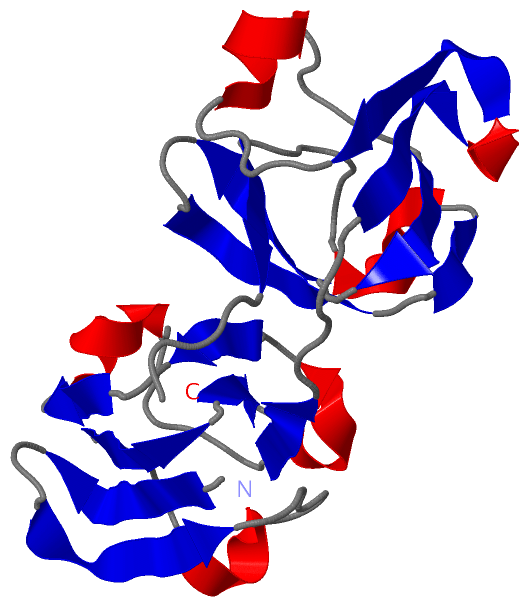
    GO:0005212    structural constituent of eye lens    The action of a molecule that contributes to the structural integrity of the lens of an eye.
biological process
    GO:0034614    cellular response to reactive oxygen species    Any process that results in a change in state or activity of a cell (in terms of movement, secretion, enzyme production, gene expression, etc.) as a result of a reactive oxygen species stimulus. Reactive oxygen species include singlet oxygen, superoxide, and oxygen free radicals.
    GO:0002088    lens development in camera-type eye    The process whose specific outcome is the progression of the lens over time, from its formation to the mature structure. The lens is a transparent structure in the eye through which light is focused onto the retina. An example of this process is found in Mus musculus.
    GO:0070306    lens fiber cell differentiation    The process in which a relatively unspecialized cell acquires specialized features of a lens fiber cell, any of the elongated, tightly packed cells that make up the bulk of the mature lens in the camera-type eye. The cytoplasm of a lens fiber cell is devoid of most intracellular organelles including the cell nucleus, and contains primarily crystallins, a group of water-soluble proteins expressed in vary large quantities.
    GO:0050896    response to stimulus    Any process that results in a change in state or activity of a cell or an organism (in terms of movement, secretion, enzyme production, gene expression, etc.) as a result of a stimulus. The process begins with detection of the stimulus and ends with a change in state or activity or the cell or organism.
    GO:0007601    visual perception    The series of events required for an organism to receive a visual stimulus, convert it to a molecular signal, and recognize and characterize the signal. Visual stimuli are detected in the form of photons and are processed to form an image.
cellular component
    GO:0005737    cytoplasm    All of the contents of a cell excluding the plasma membrane and nucleus, but including other subcellular structures.
    GO:0005634    nucleus    A membrane-bounded organelle of eukaryotic cells in which chromosomes are housed and replicated. In most cells, the nucleus contains all of the cell's chromosomes except the organellar chromosomes, and is the site of RNA synthesis and processing. In some species, or in specialized cell types, RNA metabolism or DNA replication may be absent.

 Visualization

(-) Interactive Views

Theoretical Model
  Complete Structure
    Jena3D(integrated viewing of ligand, site, SAP, PROSITE, SCOP information)
    WebMol | AstexViewer[tm]@PDBe
(Java Applets, require no local installation except for Java; loading may be slow)
    STRAP
(Java WebStart application, automatic local installation, requires Java; full application with system access!)
    RasMol
(require local installation)
    Molscript (VRML)
(requires installation of a VRML viewer; select preferred view via VRML and generate a mono or stereo PDF format file)
 
  Ligands, Modified Residues, Ions
(no "Ligands, Modified Residues, Ions" information available for 1ld0)
 
  Sites
(no "Sites" information available for 1ld0)
 
  Cis Peptide Bonds
(no "Cis Peptide Bonds" information available for 1ld0)
 

(-) Still Images

Jmol
  protein: cartoon or spacefill or dots and stick; nucleic acid: cartoon and stick; ligands: spacefill; active site: stick
Molscript
  protein, nucleic acid: cartoon; ligands: spacefill; active site: ball and stick

 Databases and Analysis Tools

(-) Databases

Access by PDB/NDB ID
  1ld0
    Family and Domain InformationProDom | SYSTERS
    General Structural InformationGlycoscienceDB | MMDB | NDB | OCA | PDB | PDBe | PDBj | PDBsum | PDBWiki | PQS | PROTEOPEDIA
    Orientation in MembranesOPM
    Protein SurfaceSURFACE
    Secondary StructureDSSP (structure derived) | HSSP (homology derived)
    Structural GenomicsGeneCensus
    Structural NeighboursCE | VAST
    Structure ClassificationCATH | Dali | SCOP
    Validation and Original DataBMRB Data View | BMRB Restraints Grid | EDS | PROCHECK | RECOORD | WHAT_CHECK
 
Access by UniProt ID/Accession number
  CRGD_HUMAN | P07320
    Comparative Protein Structure ModelsModBase
    Genomic InformationEnsembl
    Protein-protein InteractionDIP
    Sequence, Family and Domain InformationInterPro | Pfam | SMART | UniProtKB/SwissProt
 
Access by Enzyme Classificator   (EC Number)
  (no 'Enzyme Classificator' available)
    General Enzyme InformationBRENDA | EC-PDB | Enzyme | IntEnz
    PathwayKEGG | MetaCyc
 
Access by Disease Identifier   (MIM ID)
  115700
    Disease InformationOMIM
 
Access by GenAge ID
  (no 'GenAge ID' available)
    Age Related InformationGenAge

(-) Analysis Tools

Access by PDB/NDB ID
    Domain InformationXDom
    Interatomic Contacts of Structural UnitsCSU
    Ligand-protein ContactsLPC
    Protein CavitiescastP
    Sequence and Secondary StructurePDBCartoon
    Structure AlignmentSTRAP(Java WebStart application, automatic local installation, requires Java; full application with system access!)
    Structure and Sequence BrowserSTING
 
Access by UniProt ID/Accession number
  CRGD_HUMAN | P07320
    Protein Disorder PredictionDisEMBL | FoldIndex | GLOBPLOT (for more information see DisProt)

 Related Entries

(-) Entries Sharing at Least One Protein Chain (UniProt ID)

UniProtKB/Swiss-Prot
        CRGD_HUMAN | P073201h4a 1hk0 2g98 2kfb 2klj 4gr7 4jgf

(-) Related Entries Specified in the PDB File

(no "Related Entries Specified in the PDB File" available for 1LD0)