Show PDB file:   
         Plain Text   HTML   (compressed file size)
QuickSearch:   
by PDB,NDB,UniProt,PROSITE Code or Search Term(s)  
(-)Biol.Unit 1 - manually
(-)Asym.Unit - manually
(-)Asymmetric Unit
(-)Biological Unit 1
collapse expand < >
Image Biol.Unit 1 - manually
Biol.Unit 1 - manually  (Jmol Viewer)
Image Asym.Unit - manually
Asym.Unit - manually  (Jmol Viewer)
Image Asymmetric Unit
Asymmetric Unit  (Jmol Viewer)
Image Biological Unit 1
Biological Unit 1  (Jmol Viewer)

(-) Description

Title :  CRYSTAL STRUCTURE OF THE RECEPTOR-BINDING DOMAIN OF ADENOVIRUS TYPE 5 FIBER PROTEIN AT 1.7 ANGSTROMS RESOLUTION
 
Authors :  D. Xia, L. J. Henry, R. D. Gerard, J. Deisenhofer
Date :  06 Jan 95  (Deposition) - 31 Mar 95  (Release) - 24 Feb 09  (Revision)
Method :  X-RAY DIFFRACTION
Resolution :  1.70
Chains :  Asym. Unit :  A
Biol. Unit 1:  A  (3x)
Keywords :  Cell Receptor Recognition (Keyword Search: [Gene Ontology, PubMed, Web (Google))
 
Reference :  D. Xia, L. J. Henry, R. D. Gerard, J. Deisenhofer
Crystal Structure Of The Receptor-Binding Domain Of Adenovirus Type 5 Fiber Protein At 1. 7 A Resolution.
Structure V. 2 1259 1994
PubMed-ID: 7704534  |  Reference-DOI: 10.1016/S0969-2126(94)00126-X
(for further references see the PDB file header)

(-) Compounds

Molecule 1 - ADENOVIRUS TYPE 5 FIBER PROTEIN
    ChainsA
    EngineeredYES
    Expression SystemESCHERICHIA COLI
    Expression System Taxid562
    Organism ScientificHUMAN ADENOVIRUS 5
    Organism Taxid28285

 Structural Features

(-) Chains, Units

  1
Asymmetric Unit A
Biological Unit 1 (3x)A

Summary Information (see also Sequences/Alignments below)

(-) Ligands, Modified Residues, Ions  (0, 0)

(no "Ligand,Modified Residues,Ions" information available for 1KNB)

(-) Sites  (0, 0)

(no "Site" information available for 1KNB)

(-) SS Bonds  (0, 0)

(no "SS Bond" information available for 1KNB)

(-) Cis Peptide Bonds  (1, 1)

Asymmetric Unit
No.Residues
1Ala A:446 -Pro A:447

 Sequence-Structure Mapping

(-) SAPs(SNPs)/Variants  (0, 0)

(no "SAP(SNP)/Variant" information available for 1KNB)

(-) PROSITE Motifs  (0, 0)

(no "PROSITE Motif" information available for 1KNB)

(-) Exons   (0, 0)

(no "Exon" information available for 1KNB)

(-) Sequences/Alignments

Asymmetric Unit
   Reformat: Number of residues per line =  ('0' or empty: single-line sequence representation)
  Number of residues per labelling interval =   
  UniProt sequence: complete  aligned part    
   Show mapping: SCOP domains CATH domains Pfam domains Secondary structure (by author)
SAPs(SNPs) PROSITE motifs Exons
(details for a mapped element are shown in a popup box when the mouse pointer rests over it)
Chain A from PDB  Type:PROTEIN  Length:186
 aligned with SPIKE_ADE05 | P11818 from UniProtKB/Swiss-Prot  Length:581

    Alignment length:186
                                   405       415       425       435       445       455       465       475       485       495       505       515       525       535       545       555       565       575      
          SPIKE_ADE05   396 NDKLTLWTTPAPSPNCRLNAEKDAKLTLVLTKCGSQILATVSVLAVKGSLAPISGTVQSAHLIIRFDENGVLLNNSFLDPEYWNFRNGDLTEGTAYTNAVGFMPNLSAYPKSHGKTAKSNIVSQVYLNGDKTKPVTLTITLNGTQETGDTTPSAYSMSFSWDWSGHNYINEIFATSSYTFSYIAQE 581
               SCOP domains d1knba_ A: Adenovirus fiber protein knob domain                                                                                                                                            SCOP domains
               CATH domains 1knbA00 A:396-581 Adenovirus Type 5 Fiber Protein (Receptor Binding Domain)                                                                                                                CATH domains
               Pfam domains ------------------------------------------------------------------------------------------------------------------------------------------------------------------------------------------ Pfam domains
         Sec.struct. author ....eee...............eeeeeeeeee..eeeeeeeeeee..hhh........eeeeeeee.............hhh.eeee..ee........hhh..........hhh.hhheeeeeeehhh...eeeeeeee..............eeeeeee................eeeee.... Sec.struct. author
                 SAPs(SNPs) ------------------------------------------------------------------------------------------------------------------------------------------------------------------------------------------ SAPs(SNPs)
                    PROSITE ------------------------------------------------------------------------------------------------------------------------------------------------------------------------------------------ PROSITE
                 Transcript ------------------------------------------------------------------------------------------------------------------------------------------------------------------------------------------ Transcript
                 1knb A 396 NDKLTLWTTPAPSPNCRLNAEKDAKLTLVLTKCGSQILATVSVLAVKGSLAPISGTVQSAHLIIRFDENGVLLNNSFLDPEYWNFRNGDLTEGTAYTNAVGFMPNLSAYPKSHGKTAKSNIVSQVYLNGDKTKPVTLTITLNGTQETGDTTPSAYSMSFSWDWSGHNYINEIFATSSYTFSYIAQE 581
                                   405       415       425       435       445       455       465       475       485       495       505       515       525       535       545       555       565       575      

   Legend:   → Mismatch (orange background)
  - → Gap (green background, '-', border residues have a numbering label)
    → Modified Residue (blue background, lower-case, 'x' indicates undefined single-letter code, labelled with number + name)
  x → Chemical Group (purple background, 'x', labelled with number + name, e.g. ACE or NH2)
  extra numbering lines below/above indicate numbering irregularities and modified residue names etc., number ends below/above '|'

 Classification and Annotation

(-) SCOP Domains  (1, 1)

Asymmetric Unit

(-) CATH Domains  (1, 1)

Asymmetric Unit
(-)
Class: Mainly Beta (13760)

(-) Pfam Domains  (0, 0)

(no "Pfam Domain" information available for 1KNB)

(-) Gene Ontology  (10, 10)

Asymmetric Unit(hide GO term definitions)
Chain A   (SPIKE_ADE05 | P11818)
biological process
    GO:0007155    cell adhesion    The attachment of a cell, either to another cell or to an underlying substrate such as the extracellular matrix, via cell adhesion molecules.
    GO:0009405    pathogenesis    The set of specific processes that generate the ability of an organism to induce an abnormal, generally detrimental state in another organism.
    GO:0046718    viral entry into host cell    The process that occurs after viral attachment by which a virus, or viral nucleic acid, breaches the plasma membrane or cell envelope and enters the host cell. The process ends when the viral nucleic acid is released into the host cell cytoplasm.
    GO:0019058    viral life cycle    A set of processes which all viruses follow to ensure survival; includes attachment and entry of the virus particle, decoding of genome information, translation of viral mRNA by host ribosomes, genome replication, and assembly and release of viral particles containing the genome.
    GO:0016032    viral process    A multi-organism process in which a virus is a participant. The other participant is the host. Includes infection of a host cell, replication of the viral genome, and assembly of progeny virus particles. In some cases the viral genetic material may integrate into the host genome and only subsequently, under particular circumstances, 'complete' its life cycle.
    GO:0019062    virion attachment to host cell    The process by which a virion protein binds to molecules on the host cellular surface or host cell surface projection.
cellular component
    GO:0042025    host cell nucleus    A membrane-bounded organelle as it is found in the host cell in which chromosomes are housed and replicated. The host is defined as the larger of the organisms involved in a symbiotic interaction.
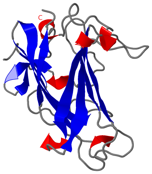
    GO:0019028    viral capsid    The protein coat that surrounds the infective nucleic acid in some virus particles. It comprises numerous regularly arranged subunits, or capsomeres.
    GO:0098022    viral capsid, fiber    A type of capsid decoration composed of fiber structures.
    GO:0019012    virion    The complete fully infectious extracellular virus particle.

 Visualization

(-) Interactive Views

Asymmetric Unit
  Complete Structure
    Jena3D(integrated viewing of ligand, site, SAP, PROSITE, SCOP information)
    WebMol | AstexViewer[tm]@PDBe
(Java Applets, require no local installation except for Java; loading may be slow)
    STRAP
(Java WebStart application, automatic local installation, requires Java; full application with system access!)
    RasMol
(require local installation)
    Molscript (VRML)
(requires installation of a VRML viewer; select preferred view via VRML and generate a mono or stereo PDF format file)
 
  Ligands, Modified Residues, Ions
(no "Ligands, Modified Residues, Ions" information available for 1knb)
 
  Sites
(no "Sites" information available for 1knb)
 
  Cis Peptide Bonds
    Ala A:446 - Pro A:447   [ RasMol ]  
 
Biological Unit
  Complete Structure
    Biological Unit 1  [ Jena3D ]

(-) Still Images

Jmol
  protein: cartoon or spacefill or dots and stick; nucleic acid: cartoon and stick; ligands: spacefill; active site: stick
Molscript
  protein, nucleic acid: cartoon; ligands: spacefill; active site: ball and stick

 Databases and Analysis Tools

(-) Databases

Access by PDB/NDB ID
  1knb
    Family and Domain InformationProDom | SYSTERS
    General Structural InformationGlycoscienceDB | MMDB | NDB | OCA | PDB | PDBe | PDBj | PDBsum | PDBWiki | PQS | PROTEOPEDIA
    Orientation in MembranesOPM
    Protein SurfaceSURFACE
    Secondary StructureDSSP (structure derived) | HSSP (homology derived)
    Structural GenomicsGeneCensus
    Structural NeighboursCE | VAST
    Structure ClassificationCATH | Dali | SCOP
    Validation and Original DataBMRB Data View | BMRB Restraints Grid | EDS | PROCHECK | RECOORD | WHAT_CHECK
 
Access by UniProt ID/Accession number
  SPIKE_ADE05 | P11818
    Comparative Protein Structure ModelsModBase
    Genomic InformationEnsembl
    Protein-protein InteractionDIP
    Sequence, Family and Domain InformationInterPro | Pfam | SMART | UniProtKB/SwissProt
 
Access by Enzyme Classificator   (EC Number)
  (no 'Enzyme Classificator' available)
    General Enzyme InformationBRENDA | EC-PDB | Enzyme | IntEnz
    PathwayKEGG | MetaCyc
 
Access by Disease Identifier   (MIM ID)
  (no 'MIM ID' available)
    Disease InformationOMIM
 
Access by GenAge ID
  (no 'GenAge ID' available)
    Age Related InformationGenAge

(-) Analysis Tools

Access by PDB/NDB ID
    Domain InformationXDom
    Interatomic Contacts of Structural UnitsCSU
    Ligand-protein ContactsLPC
    Protein CavitiescastP
    Sequence and Secondary StructurePDBCartoon
    Structure AlignmentSTRAP(Java WebStart application, automatic local installation, requires Java; full application with system access!)
    Structure and Sequence BrowserSTING
 
Access by UniProt ID/Accession number
  SPIKE_ADE05 | P11818
    Protein Disorder PredictionDisEMBL | FoldIndex | GLOBPLOT (for more information see DisProt)

 Related Entries

(-) Entries Sharing at Least One Protein Chain (UniProt ID)

UniProtKB/Swiss-Prot
        SPIKE_ADE05 | P118184atz

(-) Related Entries Specified in the PDB File

(no "Related Entries Specified in the PDB File" available for 1KNB)