|
|
|
|
 Description
|
|
Compounds
|
||||||||||||||||||||||||||||||||||||||||||||
Chains, Units
Summary Information (see also Sequences/Alignments below) |
Ligands, Modified Residues, Ions (1, 4)
NMR Structure (1, 4)
|
Sites (4, 4)
NMR Structure (4, 4)
|
SS Bonds (0, 0)| (no "SS Bond" information available for 1JFK) |
Cis Peptide Bonds (0, 0)| (no "Cis Peptide Bond" information available for 1JFK) |
SAPs(SNPs)/Variants (0, 0)| (no "SAP(SNP)/Variant" information available for 1JFK) |
PROSITE Motifs (2, 8)
NMR Structure (2, 8)
|
||||||||||||||||||||||||||||||||
Exons (0, 0)| (no "Exon" information available for 1JFK) |
Sequences/Alignments
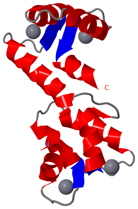
NMR StructureChain A from PDB Type:PROTEIN Length:134 aligned with CALBP_ENTHI | P38505 from UniProtKB/Swiss-Prot Length:134 Alignment length:134 10 20 30 40 50 60 70 80 90 100 110 120 130 CALBP_ENTHI 1 MAEALFKEIDVNGDGAVSYEEVKAFVSKKRAIKNEQLLQLIFKSIDADGNGEIDQNEFAKFYGSIQGQDLSDDKIGLKVLYKLMDVDGDGKLTKEEVTSFFKKHGIEKVAEQVMKADANGDGYITLEEFLEFSL 134 SCOP domains d1jfka_ A: EHCABP SCOP domains CATH domains 1jfkA01 A:1-63 EF-hand 1jfkA02 A:64-113 EF-hand --------------------- CATH domains Pfam domains efhand-1jfkA01 A:1-29 --------------------------------------------------------------------------------------------------------- Pfam domains SAPs(SNPs) -------------------------------------------------------------------------------------------------------------------------------------- SAPs(SNPs) PROSITE (1) EF_HAND_2 PDB: A:1-32 EF_HAND_2 PDB: A:33-68 ---EF_HAND_2 PDB: A:72-107 EF_HAND_2 PDB: A:108-134 PROSITE (1) PROSITE (2) ---------EF_HAND_1 -----------------------EF_HAND_1 --------------------------EF_HAND_1 -------------------EF_HAND_1 ----- PROSITE (2) Transcript -------------------------------------------------------------------------------------------------------------------------------------- Transcript 1jfk A 1 MAEALFKEIDVNGDGAVSYEEVKAFVSKKRAIKNEQLLQLIFKSIDADGNGEIDQNEFAKFYGSIQGQDLSDDKIGLKVLYKLMDVDGDGKLTKEEVTSFFKKHGIEKVAEQVMKADANGDGYITLEEFLEFSL 134 10 20 30 40 50 60 70 80 90 100 110 120 130
|
||||||||||||||||||||
SCOP Domains (1, 1)
NMR Structure
 |
CATH Domains (1, 2)
NMR Structure
|
Pfam Domains (1, 1)
NMR Structure
|
Gene Ontology (2, 2)|
NMR Structure(hide GO term definitions) Chain A (CALBP_ENTHI | P38505)
|
||||||||||||||||||
Interactive Views
|
|||||||||||||||||||||||||||||||||||||||||||||||||||||||||||||||||||||||||||||||||||||||||||||||||||||||||||||||||||||||||||||||||||||||||||
Still Images
|
||||||||||||||||
Databases
|
||||||||||||||||||||||||||||||||||||||||||||||||||||||||||||||||||||||||||||||||||||||||||||||||||||||||||||||||||||||||||||||||||||||||||||||||||||||||||||||||
Analysis Tools
|
|||||||||||||||||||||||||||||||||||||||||||||||||||||||||||||
Entries Sharing at Least One Protein Chain (UniProt ID)
Related Entries Specified in the PDB File
|
|