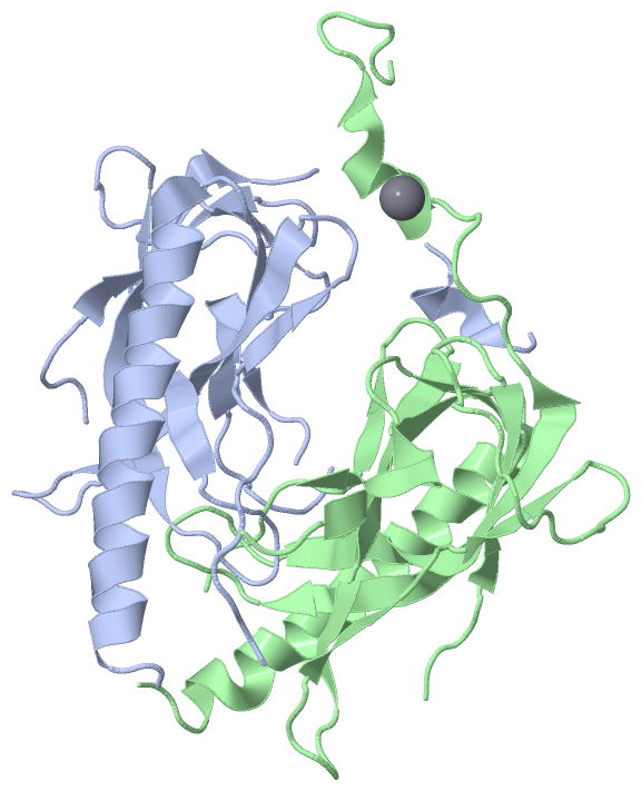
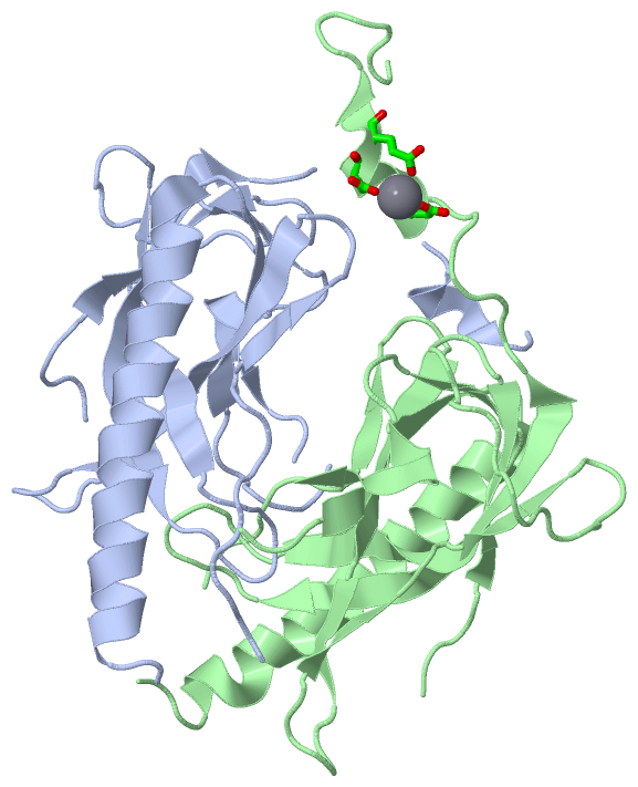
Show PDB file:   
         Plain Text   HTML   (compressed file size)
QuickSearch:   
by PDB,NDB,UniProt,PROSITE Code or Search Term(s)  
(-)Asym./Biol. Unit
(-)Asym./Biol. Unit - sites
collapse expand < >
Image Asym./Biol. Unit
Asym./Biol. Unit  (Jmol Viewer)
Image Asym./Biol. Unit - sites
Asym./Biol. Unit - sites  (Jmol Viewer)

(-) Description

Title :  CRYSTAL STRUCTURE OF GP2.5, A SINGLE-STRANDED DNA BINDING PROTEIN ENCODED BY BACTERIOPHAGE T7
 
Authors :  T. Hollis, J. M. Stattel, D. S. Walther, C. C. Richardson, T. E. Ellenberger
Date :  15 Jun 01  (Deposition) - 22 Aug 01  (Release) - 24 Feb 09  (Revision)
Method :  X-RAY DIFFRACTION
Resolution :  1.90
Chains :  Asym./Biol. Unit :  A,B
Keywords :  Ob-Fold, Beta Barrel, Dna Binding Protein (Keyword Search: [Gene Ontology, PubMed, Web (Google))
 
Reference :  T. Hollis, J. M. Stattel, D. S. Walther, C. C. Richardson, T. Ellenberger
Structure Of The Gene 2. 5 Protein, A Single-Stranded Dna Binding Protein Encoded By Bacteriophage T7.
Proc. Natl. Acad. Sci. Usa V. 98 9557 2001
PubMed-ID: 11481454  |  Reference-DOI: 10.1073/PNAS.171317698
(for further references see the PDB file header)

(-) Compounds

Molecule 1 - HELIX-DESTABILIZING PROTEIN
    ChainsA, B
    EngineeredYES
    Expression SystemESCHERICHIA COLI BL21(DE3)
    Expression System StrainBL21DE3
    Expression System Taxid469008
    Expression System Vector TypePET19B
    Organism ScientificENTEROBACTERIA PHAGE T7
    Organism Taxid10760
    SynonymGP2.5, SINGLE-STRANDED DNA-BINDING PROTEIN, SSB PROTEIN

 Structural Features

(-) Chains, Units

  12
Asymmetric/Biological Unit AB

Summary Information (see also Sequences/Alignments below)

(-) Ligands, Modified Residues, Ions  (1, 1)

Asymmetric/Biological Unit (1, 1)
No.NameCountTypeFull Name
1CA1Ligand/IonCALCIUM ION

(-) Sites  (1, 1)

Asymmetric Unit (1, 1)
No.NameEvidenceResiduesDescription
1AC1SOFTWAREASP B:189 , ASP B:192 , GLU B:193 , HOH B:590 , HOH B:612BINDING SITE FOR RESIDUE CA B 502

(-) SS Bonds  (0, 0)

(no "SS Bond" information available for 1JE5)

(-) Cis Peptide Bonds  (0, 0)

(no "Cis Peptide Bond" information available for 1JE5)

 Sequence-Structure Mapping

(-) SAPs(SNPs)/Variants  (0, 0)

(no "SAP(SNP)/Variant" information available for 1JE5)

(-) PROSITE Motifs  (0, 0)

(no "PROSITE Motif" information available for 1JE5)

(-) Exons   (0, 0)

(no "Exon" information available for 1JE5)

(-) Sequences/Alignments

Asymmetric/Biological Unit
   Reformat: Number of residues per line =  ('0' or empty: single-line sequence representation)
  Number of residues per labelling interval =   
  UniProt sequence: complete  aligned part    
   Show mapping: SCOP domains CATH domains Pfam domains Secondary structure (by author)
SAPs(SNPs) PROSITE motifs Exons
(details for a mapped element are shown in a popup box when the mouse pointer rests over it)
Chain A from PDB  Type:PROTEIN  Length:181
 aligned with DNBI_BPT7 | P03696 from UniProtKB/Swiss-Prot  Length:232

    Alignment length:197
                                    10        20        30        40        50        60        70        80        90       100       110       120       130       140       150       160       170       180       190       
            DNBI_BPT7     1 MAKKIFTSALGTAEPYAYIAKPDYGNEERGFGNPRGVYKVDLTIPNKDPRCQRMVDEIVKCHEEAYAAAVEEYEANPPAVARGKKPLKPYEGDMPFFDNGDGTTTFKFKCYASFQDKKTKETKHINLVVVDSKGKKMEDVPIIGGGSKLKVKYSLVPYKWNTAVGASVKLQLESVMLVELATFGGGEDDWADEVEEN 197
               SCOP domains d1je5a_ A: gp2.5                                                                                                                                                                                      SCOP domains
               CATH domains 1je5A00 A:1-197 Nucleic ac   id-binding proteins                                                                                                                                                      CATH domains
               Pfam domains ----------------------------------------------------------------------------------------------------------------------------------------------------------------------------------------------------- Pfam domains
         Sec.struct. author .....ee...eee....eeeeee...---.......eeeeeeeee..hhhhhhhhhhhhhhhhhhhhhhhhhhhh...-------..........eee....eeeeeeeee.eee......eee....ee.................eeeeeeeeeee........eeeeeeeeeeeee...------.hhhhh... Sec.struct. author
                 SAPs(SNPs) ----------------------------------------------------------------------------------------------------------------------------------------------------------------------------------------------------- SAPs(SNPs)
                    PROSITE ----------------------------------------------------------------------------------------------------------------------------------------------------------------------------------------------------- PROSITE
                 Transcript ----------------------------------------------------------------------------------------------------------------------------------------------------------------------------------------------------- Transcript
                 1je5 A   1 MAKKIFTSALGTAEPYAYIAKPDYGN---GFGNPRGVYKVDLTIPNKDPRCQRMVDEIVKCHEEAYAAAVEEYEANPP-------PLKPYEGDMPFFDNGDGTTTFKFKCYASFQDKKTKETKHINLVVVDSKGKKMEDVPIIGGGSKLKVKYSLVPYKWNTAVGASVKLQLESVMLVELAT------DWADEVEEN 197
                                    10        20     |  30        40        50        60        70       | -     |  90       100       110       120       130       140       150       160       170       180 |     190       
                                                    26  30                                              78      86                                                                                             182    189        

Chain B from PDB  Type:PROTEIN  Length:182
 aligned with DNBI_BPT7 | P03696 from UniProtKB/Swiss-Prot  Length:232

    Alignment length:201
                                    11        21        31        41        51        61        71        81        91       101       111       121       131       141       151       161       171       181       191       201 
            DNBI_BPT7     2 AKKIFTSALGTAEPYAYIAKPDYGNEERGFGNPRGVYKVDLTIPNKDPRCQRMVDEIVKCHEEAYAAAVEEYEANPPAVARGKKPLKPYEGDMPFFDNGDGTTTFKFKCYASFQDKKTKETKHINLVVVDSKGKKMEDVPIIGGGSKLKVKYSLVPYKWNTAVGASVKLQLESVMLVELATFGGGEDDWADEVEENGYVAS 202
               SCOP domains d1je5b_ B: gp2.5                                                                                                                                                                                          SCOP domains
               CATH domains 1je5B00 B:2-202 Nucleic          acid-binding proteins                                                                                                                                                    CATH domains
               Pfam domains --------------------------------------------------------------------------------------------------------------------------------------------------------------------------------------------------------- Pfam domains
         Sec.struct. author ....ee...eee....eeeeee.---------...eeeeeeeee..hhhhhhhhhhhhhhhhhhhhhhhhhhh....----------.......eee....eeeeeeeee.eee......eee...eee.................eeeeeeeeeee........eeeeeeeeeeeee.......hhhhhhhh........ Sec.struct. author
                 SAPs(SNPs) --------------------------------------------------------------------------------------------------------------------------------------------------------------------------------------------------------- SAPs(SNPs)
                    PROSITE --------------------------------------------------------------------------------------------------------------------------------------------------------------------------------------------------------- PROSITE
                 Transcript --------------------------------------------------------------------------------------------------------------------------------------------------------------------------------------------------------- Transcript
                 1je5 B   2 AKKIFTSALGTAEPYAYIAKPDY---------PRGVYKVDLTIPNKDPRCQRMVDEIVKCHEEAYAAAVEEYEANPP----------PYEGDMPFFDNGDGTTTFKFKCYASFQDKKTKETKHINLVVVDSKGKKMEDVPIIGGGSKLKVKYSLVPYKWNTAVGASVKLQLESVMLVELATFGGGEDDWADEVEENGYVAS 202
                                    11        21  |      -  |     41        51        61        71      |  -       |91       101       111       121       131       141       151       161       171       181       191       201 
                                                 24        34                                          78         89                                                                                                                 

   Legend:   → Mismatch (orange background)
  - → Gap (green background, '-', border residues have a numbering label)
    → Modified Residue (blue background, lower-case, 'x' indicates undefined single-letter code, labelled with number + name)
  x → Chemical Group (purple background, 'x', labelled with number + name, e.g. ACE or NH2)
  extra numbering lines below/above indicate numbering irregularities and modified residue names etc., number ends below/above '|'

 Classification and Annotation

(-) SCOP Domains  (1, 2)

Asymmetric/Biological Unit

(-) CATH Domains  (1, 2)

Asymmetric/Biological Unit
(-)
Class: Mainly Beta (13760)

(-) Pfam Domains  (0, 0)

(no "Pfam Domain" information available for 1JE5)

(-) Gene Ontology  (5, 5)

Asymmetric/Biological Unit(hide GO term definitions)
Chain A,B   (DNBI_BPT7 | P03696)
molecular function
    GO:0003677    DNA binding    Any molecular function by which a gene product interacts selectively and non-covalently with DNA (deoxyribonucleic acid).
biological process
    GO:0006310    DNA recombination    Any process in which a new genotype is formed by reassortment of genes resulting in gene combinations different from those that were present in the parents. In eukaryotes genetic recombination can occur by chromosome assortment, intrachromosomal recombination, or nonreciprocal interchromosomal recombination. Intrachromosomal recombination occurs by crossing over. In bacteria it may occur by genetic transformation, conjugation, transduction, or F-duction.
    GO:0006281    DNA repair    The process of restoring DNA after damage. Genomes are subject to damage by chemical and physical agents in the environment (e.g. UV and ionizing radiations, chemical mutagens, fungal and bacterial toxins, etc.) and by free radicals or alkylating agents endogenously generated in metabolism. DNA is also damaged because of errors during its replication. A variety of different DNA repair pathways have been reported that include direct reversal, base excision repair, nucleotide excision repair, photoreactivation, bypass, double-strand break repair pathway, and mismatch repair pathway.
    GO:0006260    DNA replication    The cellular metabolic process in which a cell duplicates one or more molecules of DNA. DNA replication begins when specific sequences, known as origins of replication, are recognized and bound by initiation proteins, and ends when the original DNA molecule has been completely duplicated and the copies topologically separated. The unit of replication usually corresponds to the genome of the cell, an organelle, or a virus. The template for replication can either be an existing DNA molecule or RNA.
    GO:0006974    cellular response to DNA damage stimulus    Any process that results in a change in state or activity of a cell (in terms of movement, secretion, enzyme production, gene expression, etc.) as a result of a stimulus indicating damage to its DNA from environmental insults or errors during metabolism.

 Visualization

(-) Interactive Views

Asymmetric/Biological Unit
  Complete Structure
    Jena3D(integrated viewing of ligand, site, SAP, PROSITE, SCOP information)
    WebMol | AstexViewer[tm]@PDBe
(Java Applets, require no local installation except for Java; loading may be slow)
    STRAP
(Java WebStart application, automatic local installation, requires Java; full application with system access!)
    RasMol
(require local installation)
    Molscript (VRML)
(requires installation of a VRML viewer; select preferred view via VRML and generate a mono or stereo PDF format file)
 
  Ligands, Modified Residues, Ions
    CA  [ RasMol | Jena3D ]  +environment [ RasMol | Jena3D ]
 
  Sites
    AC1  [ RasMol ]  +environment [ RasMol ]
 
  Cis Peptide Bonds
(no "Cis Peptide Bonds" information available for 1je5)
 

(-) Still Images

Jmol
  protein: cartoon or spacefill or dots and stick; nucleic acid: cartoon and stick; ligands: spacefill; active site: stick
Molscript
  protein, nucleic acid: cartoon; ligands: spacefill; active site: ball and stick

 Databases and Analysis Tools

(-) Databases

Access by PDB/NDB ID
  1je5
    Family and Domain InformationProDom | SYSTERS
    General Structural InformationGlycoscienceDB | MMDB | NDB | OCA | PDB | PDBe | PDBj | PDBsum | PDBWiki | PQS | PROTEOPEDIA
    Orientation in MembranesOPM
    Protein SurfaceSURFACE
    Secondary StructureDSSP (structure derived) | HSSP (homology derived)
    Structural GenomicsGeneCensus
    Structural NeighboursCE | VAST
    Structure ClassificationCATH | Dali | SCOP
    Validation and Original DataBMRB Data View | BMRB Restraints Grid | EDS | PROCHECK | RECOORD | WHAT_CHECK
 
Access by UniProt ID/Accession number
  DNBI_BPT7 | P03696
    Comparative Protein Structure ModelsModBase
    Genomic InformationEnsembl
    Protein-protein InteractionDIP
    Sequence, Family and Domain InformationInterPro | Pfam | SMART | UniProtKB/SwissProt
 
Access by Enzyme Classificator   (EC Number)
  (no 'Enzyme Classificator' available)
    General Enzyme InformationBRENDA | EC-PDB | Enzyme | IntEnz
    PathwayKEGG | MetaCyc
 
Access by Disease Identifier   (MIM ID)
  (no 'MIM ID' available)
    Disease InformationOMIM
 
Access by GenAge ID
  (no 'GenAge ID' available)
    Age Related InformationGenAge

(-) Analysis Tools

Access by PDB/NDB ID
    Domain InformationXDom
    Interatomic Contacts of Structural UnitsCSU
    Ligand-protein ContactsLPC
    Protein CavitiescastP
    Sequence and Secondary StructurePDBCartoon
    Structure AlignmentSTRAP(Java WebStart application, automatic local installation, requires Java; full application with system access!)
    Structure and Sequence BrowserSTING
 
Access by UniProt ID/Accession number
  DNBI_BPT7 | P03696
    Protein Disorder PredictionDisEMBL | FoldIndex | GLOBPLOT (for more information see DisProt)

 Related Entries

(-) Entries Sharing at Least One Protein Chain (UniProt ID)

(no "Entries Sharing at Least One Protein Chain" available for 1JE5)

(-) Related Entries Specified in the PDB File

(no "Related Entries Specified in the PDB File" available for 1JE5)