Show PDB file:   
         Plain Text   HTML   (compressed file size)
QuickSearch:   
by PDB,NDB,UniProt,PROSITE Code or Search Term(s)  
(-)NMR Structure - model 1
(-)NMR Structure - model 1, sites
(-)NMR Structure - all models
collapse expand < >
Image NMR Structure - model 1
NMR Structure - model 1  (Jmol Viewer)
Image NMR Structure - model 1, sites
NMR Structure - model 1, sites  (Jmol Viewer)
Image NMR Structure - all models
NMR Structure - all models  (Jmol Viewer)

(-) Description

Title :  UNMYRISTOYLATED GCAP-2 WITH THREE CALCIUM IONS BOUND
 
Authors :  J. B. Ames, A. M. Dizhoor, M. Ikura, K. Palczewski, L. Stryer
Date :  03 Apr 99  (Deposition) - 10 Dec 99  (Release) - 24 Feb 09  (Revision)
Method :  SOLUTION NMR
Resolution :  NOT APPLICABLE
Chains :  NMR Structure  :  A  (22x)
Keywords :  Ef-Hand, Calcium-Binding Protein, Guanylyl Cyclase Regulation, Lyase (Keyword Search: [Gene Ontology, PubMed, Web (Google))
 
Reference :  J. B. Ames, A. M. Dizhoor, M. Ikura, K. Palczewski, L. Stryer
Three-Dimensional Structure Of Guanylyl Cyclase Activating Protein-2, A Calcium-Sensitive Modulator Of Photoreceptor Guanylyl Cyclases.
J. Biol. Chem. V. 274 19329 1999
PubMed-ID: 10383444  |  Reference-DOI: 10.1074/JBC.274.27.19329
(for further references see the PDB file header)

(-) Compounds

Molecule 1 - PROTEIN (GUANYLATE CYCLASE ACTIVATING PROTEIN 2)
    CellROD
    Cellular LocationCYTOPLASM
    ChainsA
    EC Number4.6.1.2
    EngineeredYES
    Expression SystemESCHERICHIA COLI BL21
    Expression System Cell LineBL21(DE3)
    Expression System PlasmidPET11B
    Expression System StrainBL21
    Expression System Taxid511693
    Organism CommonCATTLE
    Organism ScientificBOS TAURUS
    Organism Taxid9913
    SynonymGCAP-2
    TissueRETINA

 Structural Features

(-) Chains, Units

  
NMR Structure (22x)

Summary Information (see also Sequences/Alignments below)

(-) Ligands, Modified Residues, Ions  (1, 3)

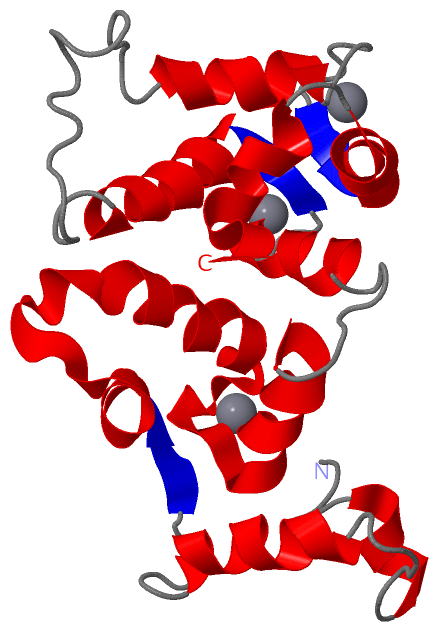
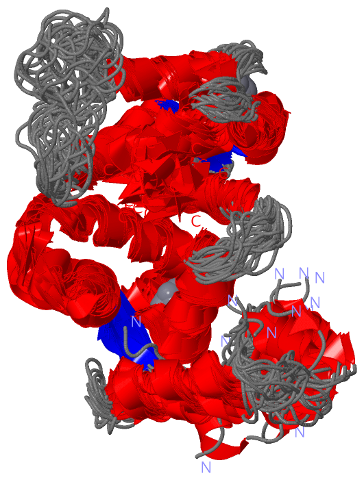
NMR Structure (1, 3)
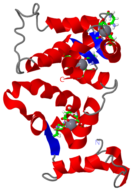
No.NameCountTypeFull Name
1CA3Ligand/IonCALCIUM ION

(-) Sites  (3, 3)

NMR Structure (3, 3)
No.NameEvidenceResiduesDescription
1AC1SOFTWAREASP A:69 , ASN A:71 , ASP A:73 , THR A:75 , GLU A:80BINDING SITE FOR RESIDUE CA A 500
2AC2SOFTWAREASP A:105 , ASP A:107 , ASN A:109 , CYS A:111 , GLU A:116BINDING SITE FOR RESIDUE CA A 501
3AC3SOFTWAREASP A:158 , ASN A:160 , ASP A:162 , GLN A:164 , GLU A:169BINDING SITE FOR RESIDUE CA A 502

(-) SS Bonds  (0, 0)

(no "SS Bond" information available for 1JBA)

(-) Cis Peptide Bonds  (0, 0)

(no "Cis Peptide Bond" information available for 1JBA)

 Sequence-Structure Mapping

(-) SAPs(SNPs)/Variants  (0, 0)

(no "SAP(SNP)/Variant" information available for 1JBA)

(-) PROSITE Motifs  (2, 6)

NMR Structure (2, 6)
 PROSITEUniProtKBPDB
No.IDACDescriptionIDLocationCountLocation
1EF_HAND_2PS50222 EF-hand calcium-binding domain profile.GUC1B_BOVIN56-91
92-127
145-180
  3A:56-91
A:92-127
A:145-180
2EF_HAND_1PS00018 EF-hand calcium-binding domain.GUC1B_BOVIN69-81
105-117
158-170
  3A:69-81
A:105-117
A:158-170

(-) Exons   (0, 0)

(no "Exon" information available for 1JBA)

(-) Sequences/Alignments

NMR Structure
   Reformat: Number of residues per line =  ('0' or empty: single-line sequence representation)
  Number of residues per labelling interval =   
  UniProt sequence: complete  aligned part    
   Show mapping: SCOP domains CATH domains Pfam domains Secondary structure (by author)
SAPs(SNPs) PROSITE motifs Exons
(details for a mapped element are shown in a popup box when the mouse pointer rests over it)
Chain A from PDB  Type:PROTEIN  Length:189
 aligned with GUC1B_BOVIN | P51177 from UniProtKB/Swiss-Prot  Length:204

    Alignment length:189
                                    11        21        31        41        51        61        71        81        91       101       111       121       131       141       151       161       171       181         
          GUC1B_BOVIN     2 GQQFSWEEAEENGAVGAADAAQLQEWYKKFLEECPSGTLFMHEFKRFFKVPDNEEATQYVEAMFRAFDTNGDNTIDFLEYVAALNLVLRGTLEHKLKWTFKIYDKDRNGCIDRQELLDIVESIYKLKKACSVEVEAEQQGKLLTPEEVVDRIFLLVDENGDGQLSLNEFVEGARRDKWVMKMLQMDLNP 190
               SCOP domains d1jbaa_ A: Guanylate cyclase activating protein 2, GCAP-2                                                                                                                                     SCOP domains
               CATH domains ---------1jbaA01 A:11-93 EF-hand                                                            1jbaA02 A:94-187 EF-hand                                                                      --- CATH domains
           Pfam domains (1) ----------------------------------------------------------------------------------------------efhand-1jbaA01 A:96-124      ------------------------------------------------------------------ Pfam domains (1)
           Pfam domains (2) ----------------------------------------------------------------------------------------------efhand-1jbaA02 A:96-124      ------------------------------------------------------------------ Pfam domains (2)
     Sec.struct. author (1) .....hhhhhhh.......hhhhhhhhhhhh......eeehhhhhhhhhhhhhhhhhhhhhhhhhhhhhhhhhhhhhhhhhhhhhhh.......hhhhhhhhh......eeehhhhhhhhhhhhhh...................hhhhhhhhhh.......eeehhhhhhhhhh..hhhhhhhhhhh. Sec.struct. author (1)
     Sec.struct. author (2) -------------------------------------------------------------------------eee----------------------------------------------------------------------------------------------------------------- Sec.struct. author (2)
                 SAPs(SNPs) --------------------------------------------------------------------------------------------------------------------------------------------------------------------------------------------- SAPs(SNPs)
                PROSITE (1) ------------------------------------------------------EF_HAND_2  PDB: A:56-91             EF_HAND_2  PDB: A:92-127            -----------------EF_HAND_2  PDB: A:145-180           ---------- PROSITE (1)
                PROSITE (2) -------------------------------------------------------------------EF_HAND_1    -----------------------EF_HAND_1    ----------------------------------------EF_HAND_1    -------------------- PROSITE (2)
                 Transcript --------------------------------------------------------------------------------------------------------------------------------------------------------------------------------------------- Transcript
                 1jba A   2 GQQFSWEEAEENGAVGAADAAQLQEWYKKFLEECPSGTLFMHEFKRFFKVPDNEEATQYVEAMFRAFDTNGDNTIDFLEYVAALNLVLRGTLEHKLKWTFKIYDKDRNGCIDRQELLDIVESIYKLKKACSVEVEAEQQGKLLTPEEVVDRIFLLVDENGDGQLSLNEFVEGARRDKWVMKMLQMDLNP 190
                                    11        21        31        41        51        61        71        81        91       101       111       121       131       141       151       161       171       181         

   Legend:   → Mismatch (orange background)
  - → Gap (green background, '-', border residues have a numbering label)
    → Modified Residue (blue background, lower-case, 'x' indicates undefined single-letter code, labelled with number + name)
  x → Chemical Group (purple background, 'x', labelled with number + name, e.g. ACE or NH2)
  extra numbering lines below/above indicate numbering irregularities and modified residue names etc., number ends below/above '|'

 Classification and Annotation

(-) SCOP Domains  (1, 1)

NMR Structure

(-) CATH Domains  (1, 2)

NMR Structure

(-) Pfam Domains  (1, 2)

NMR Structure
(-)
Clan: EF_hand (270)

(-) Gene Ontology  (6, 6)

NMR Structure(hide GO term definitions)
Chain A   (GUC1B_BOVIN | P51177)
molecular function
    GO:0005509    calcium ion binding    Interacting selectively and non-covalently with calcium ions (Ca2+).
    GO:0046872    metal ion binding    Interacting selectively and non-covalently with any metal ion.
biological process
    GO:0050896    response to stimulus    Any process that results in a change in state or activity of a cell or an organism (in terms of movement, secretion, enzyme production, gene expression, etc.) as a result of a stimulus. The process begins with detection of the stimulus and ends with a change in state or activity or the cell or organism.
    GO:0007601    visual perception    The series of events required for an organism to receive a visual stimulus, convert it to a molecular signal, and recognize and characterize the signal. Visual stimuli are detected in the form of photons and are processed to form an image.
cellular component
    GO:0016020    membrane    A lipid bilayer along with all the proteins and protein complexes embedded in it an attached to it.
    GO:0005886    plasma membrane    The membrane surrounding a cell that separates the cell from its external environment. It consists of a phospholipid bilayer and associated proteins.

 Visualization

(-) Interactive Views

NMR Structure
  Complete Structure
    Jena3D(integrated viewing of ligand, site, SAP, PROSITE, SCOP information)
    WebMol | AstexViewer[tm]@PDBe
(Java Applets, require no local installation except for Java; loading may be slow)
    STRAP
(Java WebStart application, automatic local installation, requires Java; full application with system access!)
    RasMol
(require local installation)
    Molscript (VRML)
(requires installation of a VRML viewer; select preferred view via VRML and generate a mono or stereo PDF format file)
 
  Ligands, Modified Residues, Ions
    CA  [ RasMol | Jena3D ]  +environment [ RasMol | Jena3D ]
 
  Sites
    AC1  [ RasMol ]  +environment [ RasMol ]
    AC2  [ RasMol ]  +environment [ RasMol ]
    AC3  [ RasMol ]  +environment [ RasMol ]
 
  Cis Peptide Bonds
(no "Cis Peptide Bonds" information available for 1jba)
 

(-) Still Images

Jmol
  protein: cartoon or spacefill or dots and stick; nucleic acid: cartoon and stick; ligands: spacefill; active site: stick
Molscript
  protein, nucleic acid: cartoon; ligands: spacefill; active site: ball and stick

 Databases and Analysis Tools

(-) Databases

Access by PDB/NDB ID
  1jba
    Family and Domain InformationProDom | SYSTERS
    General Structural InformationGlycoscienceDB | MMDB | NDB | OCA | PDB | PDBe | PDBj | PDBsum | PDBWiki | PQS | PROTEOPEDIA
    Orientation in MembranesOPM
    Protein SurfaceSURFACE
    Secondary StructureDSSP (structure derived) | HSSP (homology derived)
    Structural GenomicsGeneCensus
    Structural NeighboursCE | VAST
    Structure ClassificationCATH | Dali | SCOP
    Validation and Original DataBMRB Data View | BMRB Restraints Grid | EDS | PROCHECK | RECOORD | WHAT_CHECK
 
Access by UniProt ID/Accession number
  GUC1B_BOVIN | P51177
    Comparative Protein Structure ModelsModBase
    Genomic InformationEnsembl
    Protein-protein InteractionDIP
    Sequence, Family and Domain InformationInterPro | Pfam | SMART | UniProtKB/SwissProt
 
Access by Enzyme Classificator   (EC Number)
  4.6.1.2
    General Enzyme InformationBRENDA | EC-PDB | Enzyme | IntEnz
    PathwayKEGG | MetaCyc
 
Access by Disease Identifier   (MIM ID)
  (no 'MIM ID' available)
    Disease InformationOMIM
 
Access by GenAge ID
  (no 'GenAge ID' available)
    Age Related InformationGenAge

(-) Analysis Tools

Access by PDB/NDB ID
    Domain InformationXDom
    Interatomic Contacts of Structural UnitsCSU
    Ligand-protein ContactsLPC
    Protein CavitiescastP
    Sequence and Secondary StructurePDBCartoon
    Structure AlignmentSTRAP(Java WebStart application, automatic local installation, requires Java; full application with system access!)
    Structure and Sequence BrowserSTING
 
Access by UniProt ID/Accession number
  GUC1B_BOVIN | P51177
    Protein Disorder PredictionDisEMBL | FoldIndex | GLOBPLOT (for more information see DisProt)

 Related Entries

(-) Entries Sharing at Least One Protein Chain (UniProt ID)

(no "Entries Sharing at Least One Protein Chain" available for 1JBA)

(-) Related Entries Specified in the PDB File

(no "Related Entries Specified in the PDB File" available for 1JBA)