|
|
|
|
 Description
|
|
Compounds
|
||||||||||||||||||||||||||||||||||||||||||||||||||||
Chains, Units
Summary Information (see also Sequences/Alignments below) |
Ligands, Modified Residues, Ions (1, 1)
Asymmetric Unit (1, 1)
|
Sites (1, 1)
Asymmetric Unit (1, 1)
|
SS Bonds (0, 0)| (no "SS Bond" information available for 1J2Y) |
Cis Peptide Bonds (0, 0)| (no "Cis Peptide Bond" information available for 1J2Y) |
SAPs(SNPs)/Variants (0, 0)| (no "SAP(SNP)/Variant" information available for 1J2Y) |
PROSITE Motifs (1, 1)
Asymmetric Unit (1, 1)
|
||||||||||||||||||||||||||||||||||||||||||||||||
Exons (0, 0)| (no "Exon" information available for 1J2Y) |
Sequences/Alignments
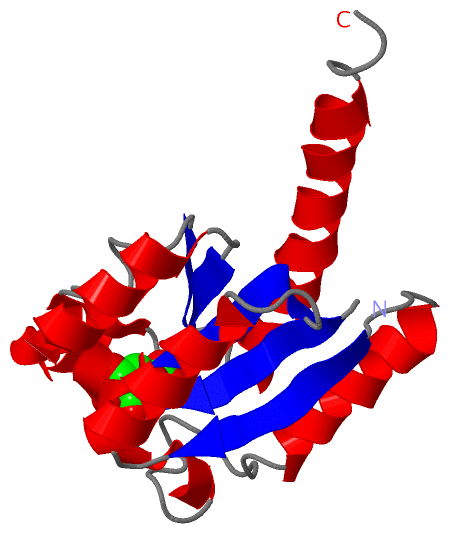
Asymmetric UnitChain A from PDB Type:PROTEIN Length:158 aligned with AROQ_HELPY | Q48255 from UniProtKB/Swiss-Prot Length:167 Alignment length:158 10 20 30 40 50 60 70 80 90 100 110 120 130 140 150 AROQ_HELPY 1 MKILVIQGPNLNMLGHRDPRLYGMVTLDQIHEIMQTFVKQGNLDVELEFFQTNFEGEIIDKIQESVGSDYEGIIINPGAFSHTSIAIADAIMLAGKPVIEVHLTNIQAREEFRKNSYTGAACGGVIMGFGPLGYNMALMAMVNILAEMKAFQEAQKNN 158 SCOP domains d1j2ya_ A: Type II 3-dehydroquinate dehydratase SCOP domains CATH domains 1j2yA00 A:1-158 [code=3.40.50.9100, no name defined] CATH domains Pfam domains -------------------------------------------------------------------------------------------------------------------------------------------------------------- Pfam domains SAPs(SNPs) -------------------------------------------------------------------------------------------------------------------------------------------------------------- SAPs(SNPs) PROSITE -----DEHYDROQUINASE_II --------------------------------------------------------------------------------------------------------------------------------------- PROSITE Transcript -------------------------------------------------------------------------------------------------------------------------------------------------------------- Transcript 1j2y A 1 MKILVIQGPNLNMLGHRDPRLYGMVTLDQIHEIMQTFVKQGNLDVELEFFQTNFEGEIIDKIQESVGSDYEGIIINPGAFSHTSIAIADAIMLAGKPVIEVHLTNIQAREEFRKNSYTGAACGGVIMGFGPLGYNMALMAMVNILAEMKAFQEAQKNN 158 10 20 30 40 50 60 70 80 90 100 110 120 130 140 150
|
||||||||||||||||||||
SCOP Domains (1, 1)
Asymmetric Unit
 |
CATH Domains (1, 1)
Asymmetric Unit
|
Pfam Domains (0, 0)| (no "Pfam Domain" information available for 1J2Y) |
Gene Ontology (5, 5)|
Asymmetric Unit(hide GO term definitions) Chain A (AROQ_HELPY | Q48255)
|
||||||||||||||||||||||||||||||||||||||||||
Interactive Views
|
||||||||||||||||||||||||||||||||||||||||||||||||||||||||||||||||||||||||||||||||||||||||||||||||||||||||||||||||||||||||||||||||||||||||
Still Images
|
||||||||||||||||
Databases
|
||||||||||||||||||||||||||||||||||||||||||||||||||||||||||||||||||||||||||||||||||||||||||||||||||||||||||||||||||||||||||||||||||||||||||||||||||||||||||||||||
Analysis Tools
|
|||||||||||||||||||||||||||||||||||||||||||||||||||||||||||||
Entries Sharing at Least One Protein Chain (UniProt ID)
Related Entries Specified in the PDB File
|
|