|
|
|
|
 Description
|
|
Compounds
|
||||||||||||||||||||||||
Chains, Units
Summary Information (see also Sequences/Alignments below) |
Ligands, Modified Residues, Ions (1, 1)
Asymmetric/Biological Unit (1, 1)
|
Sites (1, 1)
Asymmetric Unit (1, 1)
|
SS Bonds (0, 0)| (no "SS Bond" information available for 1IZD) |
Cis Peptide Bonds (2, 2)
Asymmetric/Biological Unit
|
||||||||||||
SAPs(SNPs)/Variants (0, 0)| (no "SAP(SNP)/Variant" information available for 1IZD) |
PROSITE Motifs (2, 3)
Asymmetric/Biological Unit (2, 3)
|
||||||||||||||||||||||||||||||||
Exons (0, 0)| (no "Exon" information available for 1IZD) |
Sequences/Alignments
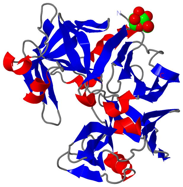
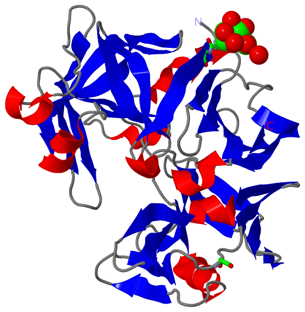
Asymmetric/Biological UnitChain A from PDB Type:PROTEIN Length:323 aligned with PEPA_ASPOZ | P0CU33 from UniProtKB/Swiss-Prot Length:390 Alignment length:323 77 87 97 107 117 127 137 147 157 167 177 187 197 207 217 227 237 247 257 267 277 287 297 307 317 327 337 347 357 367 377 387 PEPA_ASPOZ 68 AATGSVTTNPTSNDEEYITQVTVGDDTLGLDFDTGSADLWVFSSQTPSSERSGHDYYTPGSSAQKIDGATWSISYGDGSSASGDVYKDKVTVGGVSYDSQAVESAEKVSSEFTQDTANDGLLGLAFSSINTVQPTPQKTFFDNVKSSLSEPIFAVALKHNAPGVYDFGYTDSSKYTGSITYTDVDNSQGFWGFTADGYSIGSDSSSDSITGIADTGTTLLLLDDSIVDAYYEQVNGASYDSSQGGYVFPSSASLPDFSVTIGDYTATVPGEYISFADVGNGQTFGGIQSNSGIGFSIFGDVFLKSQYVVFDASGPRLGFAAQA 390 SCOP domains d1izda_ A: Acid protease SCOP domains CATH domains -----1izdA01 A:6-170 Acid Proteases 1izdA02 A:171-322 Acid Proteases - CATH domains Pfam domains ----------------------------------------------------------------------------------------------------------------------------------------------------------------------------------------------------------------------------------------------------------------------------------------------------------------------------------- Pfam domains SAPs(SNPs) ----------------------------------------------------------------------------------------------------------------------------------------------------------------------------------------------------------------------------------------------------------------------------------------------------------------------------------- SAPs(SNPs) PROSITE (1) ----------------PEPTIDASE_A1 PDB: A:17-320 UniProt: 84-387 --- PROSITE (1) PROSITE (2) -----------------------------ASP_PROTEASE-------------------------------------------------------------------------------------------------------------------------------------------------------------------------ASP_PROTEASE----------------------------------------------------------------------------------------------------- PROSITE (2) Transcript ----------------------------------------------------------------------------------------------------------------------------------------------------------------------------------------------------------------------------------------------------------------------------------------------------------------------------------- Transcript 1izd A 1 AATGSVTTNPTSNDEEYITQVTVGDDTLGLDFDTGSADLWVFSSQTPSSERSGHDYYTPGSSAQKIDGATWSISYGDGSSASGDVYKDKVTVGGVSYDSQAVESAEKVSSEFTQDTANDGLLGLAFSSINTVQPTPQKTFFDNVKSSLSEPIFAVALKHNAPGVYDFGYTDSSKYTGSITYTDVDNSQGFWGFTADGYSIGSDSSSDSITGIADTGTTLLLLDDSIVDAYYEQVNGASYDSSQGGYVFPSSASLPDFSVTIGDYTATVPGEYISFADVGNGQTFGGIQSNSGIGFSIFGDVFLKSQYVVFDASGPRLGFAAQA 323 10 20 30 40 50 60 70 80 90 100 110 120 130 140 150 160 170 180 190 200 210 220 230 240 250 260 270 280 290 300 310 320
|
||||||||||||||||||||
SCOP Domains (1, 1)
Asymmetric/Biological Unit
 |
CATH Domains (1, 2)
Asymmetric/Biological Unit
|
Pfam Domains (0, 0)| (no "Pfam Domain" information available for 1IZD) |
Gene Ontology (5, 5)|
Asymmetric/Biological Unit(hide GO term definitions) |
Interactive Views
|
||||||||||||||||||||||||||||||||||||||||||||||||||||||||||||||||||||||||||||||||||||||||||||||||||||||||||||||||||||||||||||||
Still Images
|
||||||||||||||||
Databases
|
||||||||||||||||||||||||||||||||||||||||||||||||||||||||||||||||||||||||||||||||||||||||||||||||||||||||||||||||||||||||||||||||||||||||||||||||||||||||||||||||
Analysis Tools
|
|||||||||||||||||||||||||||||||||||||||||||||||||||||||||||||
Entries Sharing at Least One Protein Chain (UniProt ID)
Related Entries Specified in the PDB File
|
|