|
|
|
|
 Description
|
|
Compounds
|
||||||||||||||||||||||||
Chains, Units
Summary Information (see also Sequences/Alignments below) |
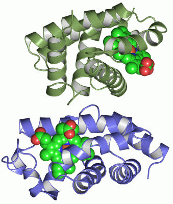
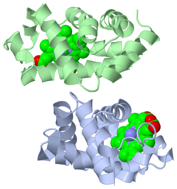
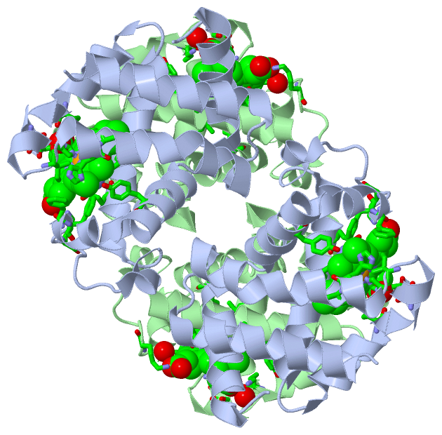
Ligands, Modified Residues, Ions (2, 4)| Asymmetric Unit (2, 4) Biological Unit 1 (2, 8) |
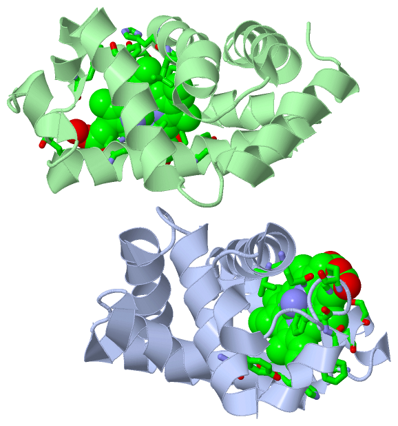
Sites (4, 4)
Asymmetric Unit (4, 4)
|
SS Bonds (0, 0)| (no "SS Bond" information available for 1ITH) |
Cis Peptide Bonds (0, 0)| (no "Cis Peptide Bond" information available for 1ITH) |
SAPs(SNPs)/Variants (0, 0)| (no "SAP(SNP)/Variant" information available for 1ITH) |
PROSITE Motifs (1, 2)
Asymmetric Unit (1, 2)
|
||||||||||||||||||||||||||||||||||||||||||||||||
Exons (0, 0)| (no "Exon" information available for 1ITH) |
Sequences/Alignments
Asymmetric UnitChain A from PDB Type:PROTEIN Length:141 aligned with HBF1_URECA | P06148 from UniProtKB/Swiss-Prot Length:142 Alignment length:141 11 21 31 41 51 61 71 81 91 101 111 121 131 141 HBF1_URECA 2 GLTTAQIKAIQDHWFLNIKGCLQAAADSIFFKYLTAYPGDLAFFHKFSSVPLYGLRSNPAYKAQTLTVINYLDKVVDALGGNAGALMKAKVPSHDAMGITPKHFGQLLKLVGGVFQEEFSADPTTVAAWGDAAGVLVAAMK 142 SCOP domains d1itha_ A: Hemoglobin SCOP domains CATH domains 1ithA00 A:1-141 Globins CATH domains Pfam domains --------------------------------------------------------------------------------------------------------------------------------------------- Pfam domains SAPs(SNPs) --------------------------------------------------------------------------------------------------------------------------------------------- SAPs(SNPs) PROSITE -GLOBIN PDB: A:2-140 UniProt: 3-141 - PROSITE Transcript --------------------------------------------------------------------------------------------------------------------------------------------- Transcript 1ith A 1 GLTAAQIKAIQDHWFLNIKGCLQAAADSIFFKYLTAYPGDLAFFHKFSSVPLYGLRSNPAYKAQTLTVINYLDKVVDALGGNAGALMKAKVPSHDAMGITPKHFGQLLKLVGGVFQEEFSADPTTVAAWGDAAGVLVAAMK 141 10 20 30 40 50 60 70 80 90 100 110 120 130 140 Chain B from PDB Type:PROTEIN Length:141 aligned with HBF1_URECA | P06148 from UniProtKB/Swiss-Prot Length:142 Alignment length:141 11 21 31 41 51 61 71 81 91 101 111 121 131 141 HBF1_URECA 2 GLTTAQIKAIQDHWFLNIKGCLQAAADSIFFKYLTAYPGDLAFFHKFSSVPLYGLRSNPAYKAQTLTVINYLDKVVDALGGNAGALMKAKVPSHDAMGITPKHFGQLLKLVGGVFQEEFSADPTTVAAWGDAAGVLVAAMK 142 SCOP domains d1ithb_ B: Hemoglobin SCOP domains CATH domains 1ithB00 B:1-141 Globins CATH domains Pfam domains --------------------------------------------------------------------------------------------------------------------------------------------- Pfam domains SAPs(SNPs) --------------------------------------------------------------------------------------------------------------------------------------------- SAPs(SNPs) PROSITE -GLOBIN PDB: B:2-140 UniProt: 3-141 - PROSITE Transcript --------------------------------------------------------------------------------------------------------------------------------------------- Transcript 1ith B 1 GLTAAQIKAIQDHWFLNIKGCLQAAADSIFFKYLTAYPGDLAFFHKFSSVPLYGLRSNPAYKAQTLTVINYLDKVVDALGGNAGALMKAKVPSHDAMGITPKHFGQLLKLVGGVFQEEFSADPTTVAAWGDAAGVLVAAMK 141 10 20 30 40 50 60 70 80 90 100 110 120 130 140
|
||||||||||||||||||||
SCOP Domains (1, 2)
Asymmetric Unit
 |
CATH Domains (1, 2)
Asymmetric Unit
|
Pfam Domains (0, 0)| (no "Pfam Domain" information available for 1ITH) |
Gene Ontology (6, 6)|
Asymmetric Unit(hide GO term definitions) Chain A,B (HBF1_URECA | P06148)
|
||||||||||||||||||||||||||||||||||||||||||||||||
Interactive Views
|
||||||||||||||||||||||||||||||||||||||||||||||||||||||||||||||||||||||||||||||||||||||||||||||||||||||||||||||||||||||||||||||||||||||||||||||||||||||||||||||||||||
Still Images
|
||||||||||||||||
Databases
|
||||||||||||||||||||||||||||||||||||||||||||||||||||||||||||||||||||||||||||||||||||||||||||||||||||||||||||||||||||||||||||||||||||||||||||||||||||||||||||||||
Analysis Tools
|
|||||||||||||||||||||||||||||||||||||||||||||||||||||||||||||
Entries Sharing at Least One Protein Chain (UniProt ID)
Related Entries Specified in the PDB File
|
|