|
|
|
|
 Description
|
|
Compounds
|
||||||||||||||||||||
Chains, Units
Summary Information (see also Sequences/Alignments below) |
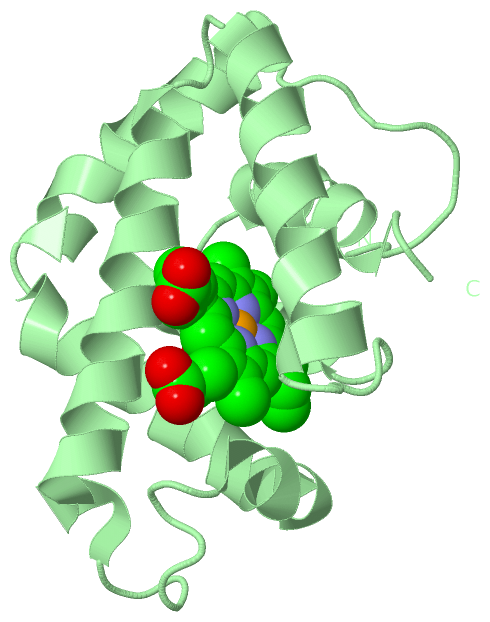
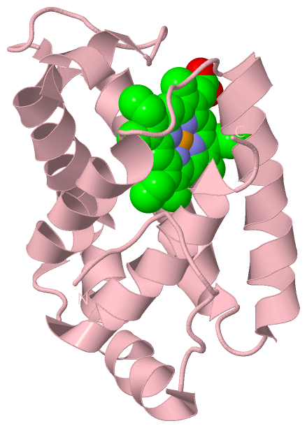
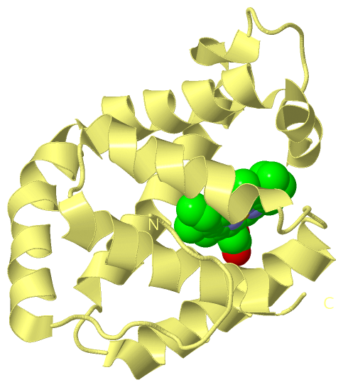
Ligands, Modified Residues, Ions (2, 8)
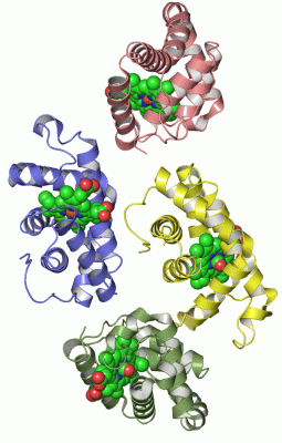
Asymmetric Unit (2, 8)
|
Sites (8, 8)
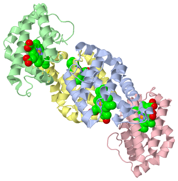
Asymmetric Unit (8, 8)
|
SS Bonds (1, 1)
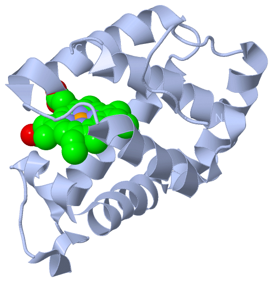
Asymmetric Unit
|
||||||||
Cis Peptide Bonds (0, 0)| (no "Cis Peptide Bond" information available for 1IT3) |
SAPs(SNPs)/Variants (0, 0)| (no "SAP(SNP)/Variant" information available for 1IT3) |
PROSITE Motifs (1, 4)
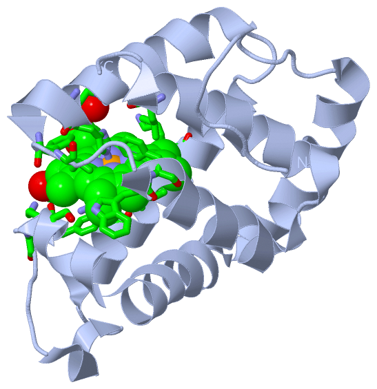
Asymmetric Unit (1, 4)
|
||||||||||||||||||||||||||||||||||||||||||||||||||||||||||||||||||||||||||||||||||||||||||||||||||||||||||||||||||||||||
Exons (0, 0)| (no "Exon" information available for 1IT3) |
Sequences/Alignments
Asymmetric UnitChain A from PDB Type:PROTEIN Length:146 aligned with GLBF1_EPTBU | Q7SID0 from UniProtKB/Swiss-Prot Length:146 Alignment length:146 10 20 30 40 50 60 70 80 90 100 110 120 130 140 GLBF1_EPTBU 1 PIIDQGPLPTLTDGDKKAINKIWPKIYKEYEQYSLNILLRFLKCFPQAQASFPKFSTKKSNLEQDPEVKHQAVVIFNKVNEIINSMDNQEEIIKSLKDLSQKHKTVFKVDSIWFKELSSIFVSTIDGGAEFEKLFSIICILLRSAY 146 SCOP domains d1it3a_ A: Hagfish hemoglobin SCOP domains CATH domains 1it3A00 A:1-146 Globins CATH domains Pfam domains -------------------------------------------------------------------------------------------------------------------------------------------------- Pfam domains SAPs(SNPs) -------------------------------------------------------------------------------------------------------------------------------------------------- SAPs(SNPs) PROSITE ----------GLOBIN PDB: A:11-146 UniProt: 11-146 PROSITE Transcript -------------------------------------------------------------------------------------------------------------------------------------------------- Transcript 1it3 A 1 PIIDQGPLPTLTDGDKKAINKIWPKIYKEYEQYSLNILLRFLKCFPQAQASFPKFSTKKSNLEQDPEVKHQAVVIFNKVNEIINSMDNQEEIIKSLKDLSQKHKTVFKVDSIWFKELSSIFVSTIDGGAEFEKLFSIICILLRSAY 146 10 20 30 40 50 60 70 80 90 100 110 120 130 140 Chain B from PDB Type:PROTEIN Length:146 aligned with GLBF1_EPTBU | Q7SID0 from UniProtKB/Swiss-Prot Length:146 Alignment length:146 10 20 30 40 50 60 70 80 90 100 110 120 130 140 GLBF1_EPTBU 1 PIIDQGPLPTLTDGDKKAINKIWPKIYKEYEQYSLNILLRFLKCFPQAQASFPKFSTKKSNLEQDPEVKHQAVVIFNKVNEIINSMDNQEEIIKSLKDLSQKHKTVFKVDSIWFKELSSIFVSTIDGGAEFEKLFSIICILLRSAY 146 SCOP domains d1it3b_ B: Hagfish hemoglobin SCOP domains CATH domains 1it3B00 B:201-346 Globins CATH domains Pfam domains -------------------------------------------------------------------------------------------------------------------------------------------------- Pfam domains SAPs(SNPs) -------------------------------------------------------------------------------------------------------------------------------------------------- SAPs(SNPs) PROSITE ----------GLOBIN PDB: B:211-346 UniProt: 11-146 PROSITE Transcript -------------------------------------------------------------------------------------------------------------------------------------------------- Transcript 1it3 B 201 PIIDQGPLPTLTDGDKKAINKIWPKIYKEYEQYSLNILLRFLKCFPQAQASFPKFSTKKSNLEQDPEVKHQAVVIFNKVNEIINSMDNQEEIIKSLKDLSQKHKTVFKVDSIWFKELSSIFVSTIDGGAEFEKLFSIICILLRSAY 346 210 220 230 240 250 260 270 280 290 300 310 320 330 340 Chain C from PDB Type:PROTEIN Length:146 aligned with GLBF1_EPTBU | Q7SID0 from UniProtKB/Swiss-Prot Length:146 Alignment length:146 10 20 30 40 50 60 70 80 90 100 110 120 130 140 GLBF1_EPTBU 1 PIIDQGPLPTLTDGDKKAINKIWPKIYKEYEQYSLNILLRFLKCFPQAQASFPKFSTKKSNLEQDPEVKHQAVVIFNKVNEIINSMDNQEEIIKSLKDLSQKHKTVFKVDSIWFKELSSIFVSTIDGGAEFEKLFSIICILLRSAY 146 SCOP domains d1it3c_ C: Hagfish hemoglobin SCOP domains CATH domains 1it3C00 C:401-546 Globins CATH domains Pfam domains -------------------------------------------------------------------------------------------------------------------------------------------------- Pfam domains SAPs(SNPs) -------------------------------------------------------------------------------------------------------------------------------------------------- SAPs(SNPs) PROSITE ----------GLOBIN PDB: C:411-546 UniProt: 11-146 PROSITE Transcript -------------------------------------------------------------------------------------------------------------------------------------------------- Transcript 1it3 C 401 PIIDQGPLPTLTDGDKKAINKIWPKIYKEYEQYSLNILLRFLKCFPQAQASFPKFSTKKSNLEQDPEVKHQAVVIFNKVNEIINSMDNQEEIIKSLKDLSQKHKTVFKVDSIWFKELSSIFVSTIDGGAEFEKLFSIICILLRSAY 546 410 420 430 440 450 460 470 480 490 500 510 520 530 540 Chain D from PDB Type:PROTEIN Length:146 aligned with GLBF1_EPTBU | Q7SID0 from UniProtKB/Swiss-Prot Length:146 Alignment length:146 10 20 30 40 50 60 70 80 90 100 110 120 130 140 GLBF1_EPTBU 1 PIIDQGPLPTLTDGDKKAINKIWPKIYKEYEQYSLNILLRFLKCFPQAQASFPKFSTKKSNLEQDPEVKHQAVVIFNKVNEIINSMDNQEEIIKSLKDLSQKHKTVFKVDSIWFKELSSIFVSTIDGGAEFEKLFSIICILLRSAY 146 SCOP domains d1it3d_ D: Hagfish hemoglobin SCOP domains CATH domains 1it3D00 D:601-746 Globins CATH domains Pfam domains -------------------------------------------------------------------------------------------------------------------------------------------------- Pfam domains SAPs(SNPs) -------------------------------------------------------------------------------------------------------------------------------------------------- SAPs(SNPs) PROSITE ----------GLOBIN PDB: D:611-746 UniProt: 11-146 PROSITE Transcript -------------------------------------------------------------------------------------------------------------------------------------------------- Transcript 1it3 D 601 PIIDQGPLPTLTDGDKKAINKIWPKIYKEYEQYSLNILLRFLKCFPQAQASFPKFSTKKSNLEQDPEVKHQAVVIFNKVNEIINSMDNQEEIIKSLKDLSQKHKTVFKVDSIWFKELSSIFVSTIDGGAEFEKLFSIICILLRSAY 746 610 620 630 640 650 660 670 680 690 700 710 720 730 740
|
||||||||||||||||||||
SCOP Domains (1, 4)
Asymmetric Unit
 |
CATH Domains (1, 4)
Asymmetric Unit
|
Pfam Domains (0, 0)| (no "Pfam Domain" information available for 1IT3) |
Gene Ontology (7, 7)|
Asymmetric Unit(hide GO term definitions) Chain A,B,C,D (GLBF1_EPTBU | Q7SID0)
|
||||||||||||||||||||||||||||||||||||||||||||||||||||||
Interactive Views
|
|||||||||||||||||||||||||||||||||||||||||||||||||||||||||||||||||||||||||||||||||||||||||||||||||||||||||||||||||||||||||||||||||||||||||||||||||||||||||||||||||||||||||||||||||||||||||||||||||||||||||||||||
Still Images
|
||||||||||||||||
Databases
|
||||||||||||||||||||||||||||||||||||||||||||||||||||||||||||||||||||||||||||||||||||||||||||||||||||||||||||||||||||||||||||||||||||||||||||||||||||||||||||||||
Analysis Tools
|
|||||||||||||||||||||||||||||||||||||||||||||||||||||||||||||
Entries Sharing at Least One Protein Chain (UniProt ID)
Related Entries Specified in the PDB File
|
|