| molecular function |
|---|
| | GO:0046875 | | ephrin receptor binding | | Interacting selectively and non-covalently with an ephrin receptor. |
| | GO:0005515 | | protein binding | | Interacting selectively and non-covalently with any protein or protein complex (a complex of two or more proteins that may include other nonprotein molecules). |
| | GO:0005102 | | receptor binding | | Interacting selectively and non-covalently with one or more specific sites on a receptor molecule, a macromolecule that undergoes combination with a hormone, neurotransmitter, drug or intracellular messenger to initiate a change in cell function. |
| biological process |
|---|
| | GO:0031295 | | T cell costimulation | | The process of providing, via surface-bound receptor-ligand pairs, a second, antigen-independent, signal in addition to that provided by the T cell receptor to augment T cell activation. |
| | GO:0001525 | | angiogenesis | | Blood vessel formation when new vessels emerge from the proliferation of pre-existing blood vessels. |
| | GO:0009887 | | animal organ morphogenesis | | Morphogenesis of an animal organ. An organ is defined as a tissue or set of tissues that work together to perform a specific function or functions. Morphogenesis is the process in which anatomical structures are generated and organized. Organs are commonly observed as visibly distinct structures, but may also exist as loosely associated clusters of cells that work together to perform a specific function or functions. |
| | GO:0007411 | | axon guidance | | The chemotaxis process that directs the migration of an axon growth cone to a specific target site in response to a combination of attractive and repulsive cues. |
| | GO:0048514 | | blood vessel morphogenesis | | The process in which the anatomical structures of blood vessels are generated and organized. The blood vessel is the vasculature carrying blood. |
| | GO:0007155 | | cell adhesion | | The attachment of a cell, either to another cell or to an underlying substrate such as the extracellular matrix, via cell adhesion molecules. |
| | GO:0030154 | | cell differentiation | | The process in which relatively unspecialized cells, e.g. embryonic or regenerative cells, acquire specialized structural and/or functional features that characterize the cells, tissues, or organs of the mature organism or some other relatively stable phase of the organism's life history. Differentiation includes the processes involved in commitment of a cell to a specific fate and its subsequent development to the mature state. |
| | GO:0016477 | | cell migration | | The controlled self-propelled movement of a cell from one site to a destination guided by molecular cues. Cell migration is a central process in the development and maintenance of multicellular organisms. |
| | GO:0002042 | | cell migration involved in sprouting angiogenesis | | The orderly movement of endothelial cells into the extracellular matrix in order to form new blood vessels involved in sprouting angiogenesis. |
| | GO:0048013 | | ephrin receptor signaling pathway | | The series of molecular signals generated as a consequence of an ephrin receptor binding to an ephrin. |
| | GO:0001945 | | lymph vessel development | | The process whose specific outcome is the progression of a lymph vessel over time, from its formation to the mature structure. |
| | GO:0007275 | | multicellular organism development | | The biological process whose specific outcome is the progression of a multicellular organism over time from an initial condition (e.g. a zygote or a young adult) to a later condition (e.g. a multicellular animal or an aged adult). |
| | GO:0010839 | | negative regulation of keratinocyte proliferation | | Any process that decreases the rate, frequency or extent of keratinocyte proliferation. Keratinocyte proliferation is the multiplication or reproduction of keratinocytes, resulting in the expansion of a cell population. |
| | GO:0072178 | | nephric duct morphogenesis | | The process in which the anatomical structures of the nephric duct are generated and organized. A nephric duct is a tube that drains a primitive kidney. |
| | GO:0007399 | | nervous system development | | The process whose specific outcome is the progression of nervous tissue over time, from its formation to its mature state. |
| | GO:1903849 | | positive regulation of aorta morphogenesis | | Any process that activates or increases the frequency, rate or extent of aorta morphogenesis. |
| | GO:2000727 | | positive regulation of cardiac muscle cell differentiation | | Any process that activates or increases the frequency, rate or extent of cardiac muscle cell differentiation. |
| | GO:0050920 | | regulation of chemotaxis | | Any process that modulates the frequency, rate or extent of the directed movement of a motile cell or organism in response to a specific chemical concentration gradient. |
| | GO:0048845 | | venous blood vessel morphogenesis | | The process in which the anatomical structures of venous blood vessels are generated and organized. Veins are blood vessels that transport blood from the body and its organs to the heart. |
| cellular component |
|---|
| | GO:0005925 | | focal adhesion | | Small region on the surface of a cell that anchors the cell to the extracellular matrix and that forms a point of termination of actin filaments. |
| | GO:0016021 | | integral component of membrane | | The component of a membrane consisting of the gene products and protein complexes having at least some part of their peptide sequence embedded in the hydrophobic region of the membrane. |
| | GO:0016020 | | membrane | | A lipid bilayer along with all the proteins and protein complexes embedded in it an attached to it. |
| | GO:0005886 | | plasma membrane | | The membrane surrounding a cell that separates the cell from its external environment. It consists of a phospholipid bilayer and associated proteins. |
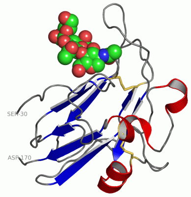
 Description
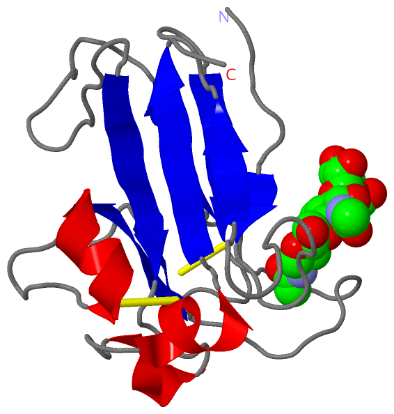