|
|
|
|
 Description
|
|
Compounds
|
||||||||||||||||||||||||||||||||||||||||||||||||||||
Chains, Units
Summary Information (see also Sequences/Alignments below) |
Ligands, Modified Residues, Ions (1, 1)
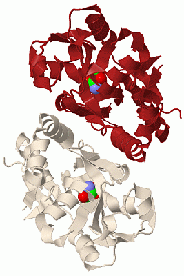
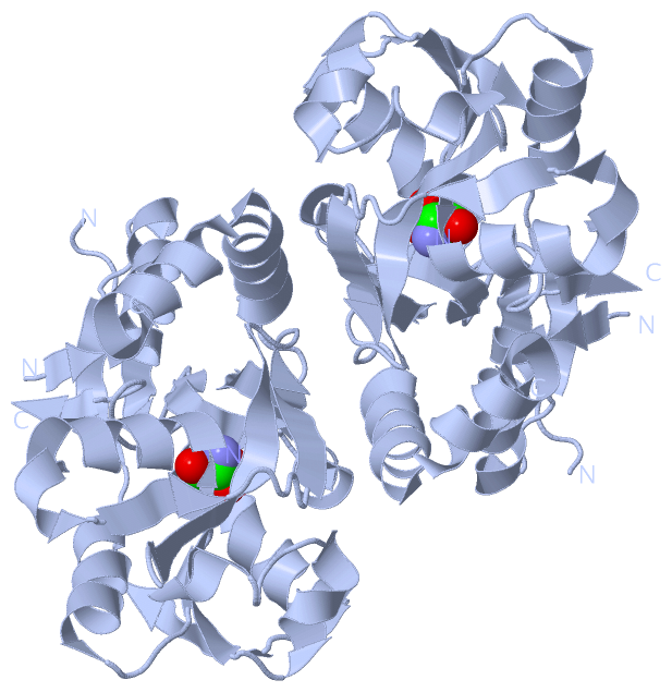
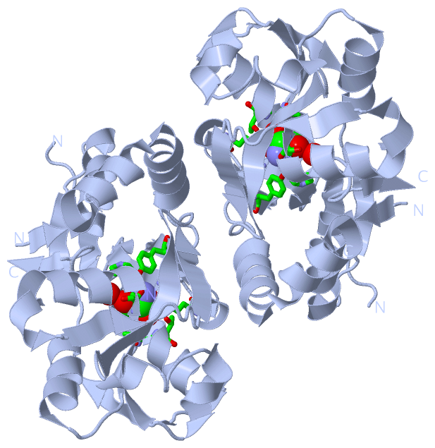
Asymmetric Unit (1, 1)
|
Sites (1, 1)
Asymmetric Unit (1, 1)
|
SS Bonds (0, 0)| (no "SS Bond" information available for 1IIT) |
Cis Peptide Bonds (1, 1)
Asymmetric Unit
|
||||||||
SAPs(SNPs)/Variants (0, 0)| (no "SAP(SNP)/Variant" information available for 1IIT) |
PROSITE Motifs (0, 0)| (no "PROSITE Motif" information available for 1IIT) |
Exons (0, 0)| (no "Exon" information available for 1IIT) |
Sequences/Alignments
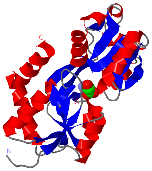
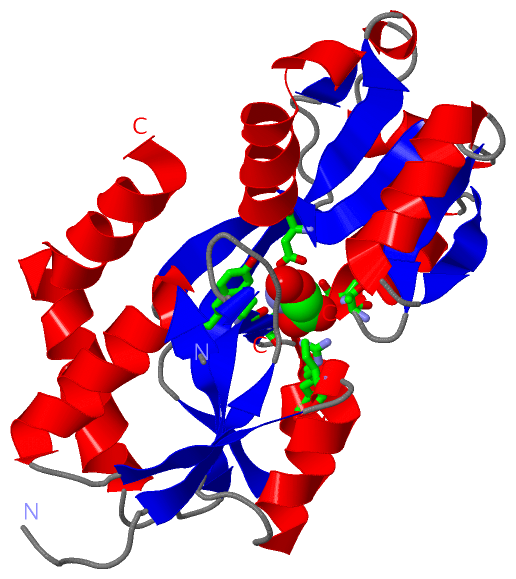
Asymmetric UnitChain A from PDB Type:PROTEIN Length:222 aligned with P73797_SYNY3 | P73797 from UniProtKB/TrEMBL Length:397 Alignment length:361 44 54 64 74 84 94 104 114 124 134 144 154 164 174 184 194 204 214 224 234 244 254 264 274 284 294 304 314 324 334 344 354 364 374 384 394 P73797_SYNY3 35 GVETVDSQTLKVGVVGNPPFVFYGEGKNAAFTGISLDVWRAVAESQKWNSEYVRQNSISAGITAVAEGELDILIGPISVTPERAAIEGITFTQPYFSSGIGLLIPGKPVSLWERFSPFFGIAALSSAGVLTLLLFLVGNLIWLAEHRKNPEQFSPHYPEGVQNGMWFALVTLTTVGYGDRSPRTKLGQLVAGVWMLVALLSFSSITAGLASAFSTALSEASATPLFRSVGDLKNKEVAVVRDTTAVDWANFYQADVRETNNLTAAITLLQKKQVEAVMFDRPALIYYTRQNPNLNLEVTEIRVSLEPYGFVLKENSPLQKTINVEMLNLLYSRVIAEFTERWLGPGIEENQDLLPQNIGES 395 SCOP domains d 1iita_ A: Glutamate receptor ligand binding core SCOP domains CATH domains 1 iitA01 A:1-94,A:188 -225 Periplasmic binding protein-like II 1iitA02 A:95-187 Periplasmic binding protein-like II 1iitA01 A:1-94,A:188-225 - - CATH domains Pfam domains ------------------------------------------------------------------------------------------------------------------------------------------------------------------------------------------------------------------------------------------------------------------------------------------------------------------------------------------------------------------------- Pfam domains
|
||||||||||||||||||||
SCOP Domains (1, 1)
Asymmetric Unit
 |
CATH Domains (1, 2)
Asymmetric Unit
|
Pfam Domains (0, 0)| (no "Pfam Domain" information available for 1IIT) |
Gene Ontology (4, 4)|
Asymmetric Unit(hide GO term definitions) Chain A (P73797_SYNY3 | P73797)
|
||||||||||||||||||||||||||||||||||||||||||
Interactive Views
|
|||||||||||||||||||||||||||||||||||||||||||||||||||||||||||||||||||||||||||||||||||||||||||||||||||||||||||||||||||||||||||||||||||||||||
Still Images
|
||||||||||||||||
Databases
|
||||||||||||||||||||||||||||||||||||||||||||||||||||||||||||||||||||||||||||||||||||||||||||||||||||||||||||||||||||||||||||||||||||||||||||||||||||||||||||||||
Analysis Tools
|
|||||||||||||||||||||||||||||||||||||||||||||||||||||||||||||
Entries Sharing at Least One Protein Chain (UniProt ID)
Related Entries Specified in the PDB File
|
|