|
|
|
|
 Description
|
|
Compounds
|
||||||||||||||||||||||||||||||||||||
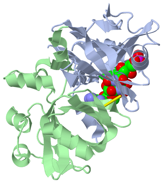
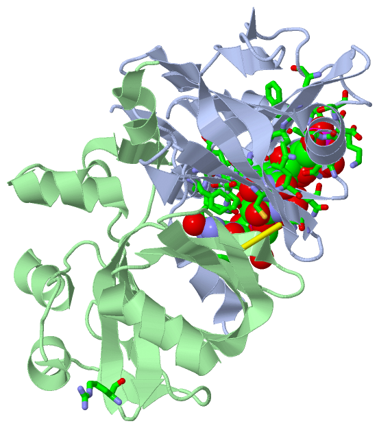
Chains, Units
Summary Information (see also Sequences/Alignments below) |
Ligands, Modified Residues, Ions (2, 2)| Asymmetric/Biological Unit (2, 2) |
Sites (2, 2)
Asymmetric Unit (2, 2)
|
SS Bonds (1, 1)
Asymmetric/Biological Unit
|
||||||||
Cis Peptide Bonds (2, 2)
Asymmetric/Biological Unit
|
||||||||||||
SAPs(SNPs)/Variants (0, 0)| (no "SAP(SNP)/Variant" information available for 1I0S) |
PROSITE Motifs (0, 0)| (no "PROSITE Motif" information available for 1I0S) |
Exons (0, 0)| (no "Exon" information available for 1I0S) |
Sequences/Alignments
Asymmetric/Biological UnitChain A from PDB Type:PROTEIN Length:161 aligned with FERCR_ARCFU | O29428 from UniProtKB/Swiss-Prot Length:169 Alignment length:161 10 20 30 40 50 60 70 80 90 100 110 120 130 140 150 160 FERCR_ARCFU 1 MDVEAFYKISYGLYIVTSESNGRKCGQIANTVFQLTSKPVQIAVCLNKENDTHNAVKESGAFGVSVLELETPMEFIGRFGFRKSSEFEKFDGVEYKTGKTGVPLVTQHAVAVIEAKVVKECDVGTHTLFVGEAVDAEVLKDAEVLTYADYHLMKKGKTPRT 161 SCOP domains d1i0sa_ A: Ferric reductase SCOP domains CATH domains 1i0sA00 A:1-161 Electron Transport, Fmn-binding Protein; Chain A CATH domains Pfam domains ----------------------------------------------------------------------------------------------------------------------------------------------------------------- Pfam domains SAPs(SNPs) ----------------------------------------------------------------------------------------------------------------------------------------------------------------- SAPs(SNPs) PROSITE ----------------------------------------------------------------------------------------------------------------------------------------------------------------- PROSITE Transcript ----------------------------------------------------------------------------------------------------------------------------------------------------------------- Transcript 1i0s A 1 MDVEAFYKISYGLYIVTSESNGRKCGQIANTVFQLTSKPVQIAVCLNKENDTHNAVKESGAFGVSVLELETPMEFIGRFGFRKSSEFEKFDGVEYKTGKTGVPLVTQHAVAVIEAKVVKECDVGTHTLFVGEAVDAEVLKDAEVLTYADYHLMKKGKTPRT 161 10 20 30 40 50 60 70 80 90 100 110 120 130 140 150 160 Chain B from PDB Type:PROTEIN Length:168 aligned with FERCR_ARCFU | O29428 from UniProtKB/Swiss-Prot Length:169 Alignment length:168 10 20 30 40 50 60 70 80 90 100 110 120 130 140 150 160 FERCR_ARCFU 1 MDVEAFYKISYGLYIVTSESNGRKCGQIANTVFQLTSKPVQIAVCLNKENDTHNAVKESGAFGVSVLELETPMEFIGRFGFRKSSEFEKFDGVEYKTGKTGVPLVTQHAVAVIEAKVVKECDVGTHTLFVGEAVDAEVLKDAEVLTYADYHLMKKGKTPRTATVYFES 168 SCOP domains d1i0sb_ B: Ferric reductase SCOP domains CATH domains 1i0sB00 B:1-168 Electron Transport, Fmn-binding Protein; Chain A CATH domains Pfam domains ------------------------------------------------------------------------------------------------------------------------------------------------------------------------ Pfam domains SAPs(SNPs) ------------------------------------------------------------------------------------------------------------------------------------------------------------------------ SAPs(SNPs) PROSITE ------------------------------------------------------------------------------------------------------------------------------------------------------------------------ PROSITE Transcript ------------------------------------------------------------------------------------------------------------------------------------------------------------------------ Transcript 1i0s B 1 MDVEAFYKISYGLYIVTSESNGRKCGQIANTVFQLTSKPVQIAVCLNKENDTHNAVKESGAFGVSVLELETPMEFIGRFGFRKSSEFEKFDGVEYKTGKTGVPLVTQHAVAVIEAKVVKECDVGTHTLFVGEAVDAEVLKDAEVLTYADYHLMKKGKTPRTATVYFES 168 10 20 30 40 50 60 70 80 90 100 110 120 130 140 150 160
|
||||||||||||||||||||
SCOP Domains (1, 2)
Asymmetric/Biological Unit
 |
CATH Domains (1, 2)
Asymmetric/Biological Unit
|
Pfam Domains (0, 0)| (no "Pfam Domain" information available for 1I0S) |
Gene Ontology (6, 6)|
Asymmetric/Biological Unit(hide GO term definitions) Chain A,B (FERCR_ARCFU | O29428)
|
||||||||||||||||||||||||||||||||||||||||||||||||
Interactive Views
|
||||||||||||||||||||||||||||||||||||||||||||||||||||||||||||||||||||||||||||||||||||||||||||||||||||||||||||||||||||||||||||||||||||||||||||
Still Images
|
||||||||||||||||
Databases
|
||||||||||||||||||||||||||||||||||||||||||||||||||||||||||||||||||||||||||||||||||||||||||||||||||||||||||||||||||||||||||||||||||||||||||||||||||||||||||||||||
Analysis Tools
|
|||||||||||||||||||||||||||||||||||||||||||||||||||||||||||||
Entries Sharing at Least One Protein Chain (UniProt ID)
Related Entries Specified in the PDB File
|
|