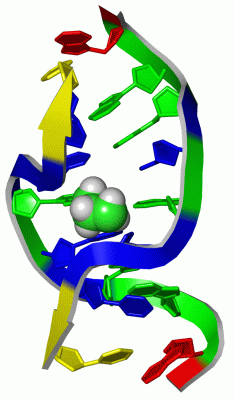
Show PDB file:   
         Plain Text   HTML   (compressed file size)
QuickSearch:   
by PDB,NDB,UniProt,PROSITE Code or Search Term(s)  
(-)NMR Structure - manually
(-)NMR Structure - model 1
(-)NMR Structure - model 1, sites
(-)NMR Structure - all models
collapse expand < >
Image NMR Structure - manually
NMR Structure - manually  (Jmol Viewer)
Image NMR Structure - model 1
NMR Structure - model 1  (Jmol Viewer)
Image NMR Structure - model 1, sites
NMR Structure - model 1, sites  (Jmol Viewer)
Image NMR Structure - all models
NMR Structure - all models  (Jmol Viewer)

(-) Description

Title :  SOLUTION NMR STRUCTURE OF SELF-COMPLEMENTARY DUPLEX 5'-D(AGGCG*CCT)2 CONTAINING A TRIMETHYLENE CROSSLINK AT THE N2 POSITION OF G*. MODEL OF A MALONDIALDEHYDE CROSSLINK
 
Authors :  P. A. Dooley, D. Tsarouhtsis, G. A. Korbel, L. V. Nechev, J. Shearer, I. S. Zegar, C. M. Harris, M. P. Stone, T. M. Harris
Date :  23 Jan 01  (Deposition) - 07 Feb 01  (Release) - 24 Feb 09  (Revision)
Method :  SOLUTION NMR
Resolution :  NOT APPLICABLE
Chains :  NMR Structure  :  A,B
Keywords :  Dna Duplex, Interstrand Crosslink (Keyword Search: [Gene Ontology, PubMed, Web (Google))
 
Reference :  P. A. Dooley, D. Tsarouhtsis, G. A. Korbel, L. V. Nechev, J. Shearer, I. S. Zegar, C. M. Harris, M. P. Stone, T. M. Harris
Structural Studies Of An Oligodeoxynucleotide Containing A Trimethylene Interstrand Cross-Link In A 5'-(Cpg) Motif: Model Of A Malondialdehyde Cross-Link.
J. Am. Chem. Soc. V. 123 1730 2001
PubMed-ID: 11456774  |  Reference-DOI: 10.1021/JA003163W
(for further references see the PDB file header)

(-) Compounds

Molecule 1 - DNA (5'-D(*AP*GP*GP*CP*GP*CP*CP*T)-3')
    ChainsA, B
    EngineeredYES
    SyntheticYES

 Structural Features

(-) Chains, Units

  
NMR Structure 

Summary Information (see also Sequences/Alignments below)

(-) Ligands, Modified Residues, Ions  (1, 1)

NMR Structure (1, 1)
No.NameCountTypeFull Name
1TME1Ligand/IonPROPANE

(-) Sites  (1, 1)

NMR Structure (1, 1)
No.NameEvidenceResiduesDescription
1AC1SOFTWAREDG A:5 , DC A:6 , DG B:13 , DC B:14BINDING SITE FOR RESIDUE TME A 9

(-) SS Bonds  (0, 0)

(no "SS Bond" information available for 1HZ2)

(-) Cis Peptide Bonds  (0, 0)

(no "Cis Peptide Bond" information available for 1HZ2)

 Sequence-Structure Mapping

(-) SAPs(SNPs)/Variants  (0, 0)

(no "SAP(SNP)/Variant" information available for 1HZ2)

(-) PROSITE Motifs  (0, 0)

(no "PROSITE Motif" information available for 1HZ2)

(-) Exons   (0, 0)

(no "Exon" information available for 1HZ2)

(-) Sequences/Alignments

NMR Structure
   Reformat: Number of residues per line =  ('0' or empty: single-line sequence representation)
  Number of residues per labelling interval =   
  UniProt sequence: complete  aligned part    
   Show mapping: SCOP domains CATH domains Pfam domains Secondary structure (by author)
SAPs(SNPs) PROSITE motifs Exons
(details for a mapped element are shown in a popup box when the mouse pointer rests over it)
Chain A from PDB  Type:DNA  Length:8
                                       
                  1hz2 A  1 AGGCGCCT  8

Chain B from PDB  Type:DNA  Length:8
                                       
                  1hz2 B  9 AGGCGCCT 16

   Legend:   → Mismatch (orange background)
  - → Gap (green background, '-', border residues have a numbering label)
    → Modified Residue (blue background, lower-case, 'x' indicates undefined single-letter code, labelled with number + name)
  x → Chemical Group (purple background, 'x', labelled with number + name, e.g. ACE or NH2)
  extra numbering lines below/above indicate numbering irregularities and modified residue names etc., number ends below/above '|'

 Classification and Annotation

(-) SCOP Domains  (0, 0)

(no "SCOP Domain" information available for 1HZ2)

(-) CATH Domains  (0, 0)

(no "CATH Domain" information available for 1HZ2)

(-) Pfam Domains  (0, 0)

(no "Pfam Domain" information available for 1HZ2)

(-) Gene Ontology  (0, 0)

NMR Structure(hide GO term definitions)
    (no "Gene Ontology" information available for 1HZ2)

 Visualization

(-) Interactive Views

NMR Structure
  Complete Structure
    Jena3D(integrated viewing of ligand, site, SAP, PROSITE, SCOP information)
    WebMol | AstexViewer[tm]@PDBe
(Java Applets, require no local installation except for Java; loading may be slow)
    STRAP
(Java WebStart application, automatic local installation, requires Java; full application with system access!)
    RasMol
(require local installation)
    Molscript (VRML)
(requires installation of a VRML viewer; select preferred view via VRML and generate a mono or stereo PDF format file)
 
  Ligands, Modified Residues, Ions
    TME  [ RasMol | Jena3D ]  +environment [ RasMol | Jena3D ]
 
  Sites
    AC1  [ RasMol ]  +environment [ RasMol ]
 
  Cis Peptide Bonds
(no "Cis Peptide Bonds" information available for 1hz2)
 

(-) Still Images

Jmol
  protein: cartoon or spacefill or dots and stick; nucleic acid: cartoon and stick; ligands: spacefill; active site: stick
Molscript
  protein, nucleic acid: cartoon; ligands: spacefill; active site: ball and stick

 Databases and Analysis Tools

(-) Databases

Access by PDB/NDB ID
  1hz2
    Family and Domain InformationProDom | SYSTERS
    General Structural InformationGlycoscienceDB | MMDB | NDB | OCA | PDB | PDBe | PDBj | PDBsum | PDBWiki | PQS | PROTEOPEDIA
    Orientation in MembranesOPM
    Protein SurfaceSURFACE
    Secondary StructureDSSP | HSSP
    Structural GenomicsGeneCensus
    Structural NeighboursCE | VAST
    Structure ClassificationCATH | Dali | SCOP
    Validation and Original DataBMRB Data View | BMRB Restraints Grid | EDS | PROCHECK | RECOORD | WHAT_CHECK
 
Access by UniProt ID/Accession number
  (no 'UniProt ID/Accession number' available) |
    Comparative Protein Structure ModelsModBase
    Genomic InformationEnsembl
    Protein-protein InteractionDIP
    Sequence, Family and Domain InformationInterPro | Pfam | SMART | UniProt
 
Access by Enzyme Classificator   (EC Number)
  (no 'Enzyme Classificator' available)
    General Enzyme InformationBRENDA | EC-PDB | Enzyme | IntEnz
    PathwayKEGG | MetaCyc
 
Access by Disease Identifier   (MIM ID)
  (no 'MIM ID' available)
    Disease InformationOMIM
 
Access by GenAge ID
  (no 'GenAge ID' available)
    Age Related InformationGenAge

(-) Analysis Tools

Access by PDB/NDB ID
    Domain InformationXDom
    Interatomic Contacts of Structural UnitsCSU
    Ligand-protein ContactsLPC
    Protein CavitiescastP
    Sequence and Secondary StructurePDBCartoon
    Structure AlignmentSTRAP(Java WebStart application, automatic local installation, requires Java; full application with system access!)
    Structure and Sequence BrowserSTING
 
Access by UniProt ID/Accession number
  (no 'UniProt ID/Accession number' available) |
    Protein Disorder PredictionDisEMBL | FoldIndex | GLOBPLOT

 Related Entries

(-) Entries Sharing at Least One Protein Chain (UniProt ID)

(no "Entries Sharing at Least One Protein Chain" available for 1HZ2)

(-) Related Entries Specified in the PDB File

(no "Related Entries Specified in the PDB File" available for 1HZ2)