Show PDB file:   
         Plain Text   HTML   (compressed file size)
QuickSearch:   
by PDB,NDB,UniProt,PROSITE Code or Search Term(s)  
(-)Asym./Biol. Unit
(-)Asym./Biol. Unit - sites
collapse expand < >
Image Asym./Biol. Unit
Asym./Biol. Unit  (Jmol Viewer)
Image Asym./Biol. Unit - sites
Asym./Biol. Unit - sites  (Jmol Viewer)

(-) Description

Title :  CRYSTAL STRUCTURE OF ALGINATE LYASE A1-III COMPLEXED WITH TRISACCHARIDE PRODUCT.
 
Authors :  H. -J. Yoon, W. Hashimoto, O. Miyake, K. Murata, B. Mikami
Date :  08 Jan 01  (Deposition) - 02 May 01  (Release) - 13 Jul 11  (Revision)
Method :  X-RAY DIFFRACTION
Resolution :  2.00
Chains :  Asym./Biol. Unit :  A
Keywords :  Alginate Lyase, Trisaccharide Complex, Alpha Barrel, Lyase (Keyword Search: [Gene Ontology, PubMed, Web (Google))
 
Reference :  H. J. Yoon, W. Hashimoto, O. Miyake, K. Murata, B. Mikami
Crystal Structure Of Alginate Lyase A1-Iii Complexed With Trisaccharide Product At 2. 0 A Resolution.
J. Mol. Biol. V. 307 9 2001
PubMed-ID: 11243798  |  Reference-DOI: 10.1006/JMBI.2000.4509
(for further references see the PDB file header)

(-) Compounds

Molecule 1 - ALGINATE LYASE
    ChainsA
    EC Number4.2.2.3
    EngineeredYES
    Expression SystemBACILLUS SUBTILIS
    Expression System PlasmidPISA412
    Expression System Taxid1423
    Expression System Vector TypePLASMID
    FragmentRESIDUE 52-402
    Organism ScientificSPHINGOMONAS SP.
    Organism Taxid28214

 Structural Features

(-) Chains, Units

  1
Asymmetric/Biological Unit A

Summary Information (see also Sequences/Alignments below)
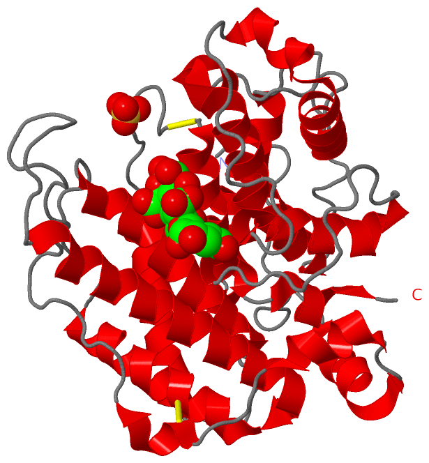
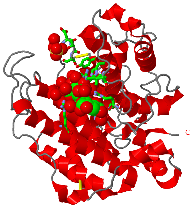
(-) Ligands, Modified Residues, Ions  (4, 4)

Asymmetric/Biological Unit (4, 4)
No.NameCountTypeFull Name
1GCU1Ligand/IonD-GLUCURONIC ACID
2MAV1Ligand/IonALPHA-D-MANNOPYRANURONIC ACID
3MAW1Ligand/Ion4-DEOXY-ALPHA-L-ERYTHRO-HEX-4-ENOPYRANURONIC ACID
4SO41Ligand/IonSULFATE ION

(-) Sites  (4, 4)

Asymmetric Unit (4, 4)
No.NameEvidenceResiduesDescription
1AC1SOFTWAREARG A:312 , GLY A:313 , MAV A:402 , HOH A:636 , HOH A:643 , HOH A:688BINDING SITE FOR RESIDUE MAW A 401
2AC2SOFTWAREARG A:306 , ARG A:312 , MAW A:401 , GCU A:403 , HOH A:634 , HOH A:635 , HOH A:636 , HOH A:744BINDING SITE FOR RESIDUE MAV A 402
3AC3SOFTWAREARG A:88 , TRP A:141 , HIS A:245 , TYR A:246 , TYR A:249 , ARG A:342 , MAV A:402 , HOH A:635 , HOH A:640 , HOH A:641 , HOH A:655 , HOH A:717BINDING SITE FOR RESIDUE GCU A 403
4AC4SOFTWAREGLY A:184 , ASP A:185 , THR A:186 , SER A:187 , HOH A:706BINDING SITE FOR RESIDUE SO4 A 801

(-) SS Bonds  (2, 2)

Asymmetric/Biological Unit
No.Residues
1A:49 -A:112
2A:188 -A:189

(-) Cis Peptide Bonds  (0, 0)

(no "Cis Peptide Bond" information available for 1HV6)

 Sequence-Structure Mapping

(-) SAPs(SNPs)/Variants  (0, 0)

(no "SAP(SNP)/Variant" information available for 1HV6)

(-) PROSITE Motifs  (0, 0)

(no "PROSITE Motif" information available for 1HV6)

(-) Exons   (0, 0)

(no "Exon" information available for 1HV6)

(-) Sequences/Alignments

Asymmetric/Biological Unit
   Reformat: Number of residues per line =  ('0' or empty: single-line sequence representation)
  Number of residues per labelling interval =   
  UniProt sequence: complete  aligned part    
   Show mapping: SCOP domains CATH domains Pfam domains Secondary structure (by author)
SAPs(SNPs) PROSITE motifs Exons
(details for a mapped element are shown in a popup box when the mouse pointer rests over it)
Chain A from PDB  Type:PROTEIN  Length:351
 aligned with Q9KWU1_SPHSX | Q9KWU1 from UniProtKB/TrEMBL  Length:641

    Alignment length:351
                                    61        71        81        91       101       111       121       131       141       151       161       171       181       191       201       211       221       231       241       251       261       271       281       291       301       311       321       331       341       351       361       371       381       391       401 
         Q9KWU1_SPHSX    52 QAHPFDQAVVKDPTASYVDVKARRTFLQSGQLDDRLKAALPKEYDCTTEATPNPQQGEMVIPRRYLSGNHGPVNPDYEPVVTLYRDFEKISATLGNLYVATGKPVYATCLLNMLDKWAKADALLNYDPKSQSWYQVEWSAATAAFALSTMMAEPNVDTAQRERVVKWLNRVARHQTSFPGGDTSCCNNHSYWRGQEATIIGVISKDDELFRWGLGRYVQAMGLINEDGSFVHEMTRHEQSLHYQNYAMLPLTMIAETASRQGIDLYAYKENGRDIHSARKFVFAAVKNPDLIKKYASEPQDTRAFKPGRGDLNWIEYQRARFGFADELGFMTVPIFDPRTGGSGTLLAYKP 402
               SCOP domains d1hv6a_ A: Alginate lyase A1-III                                                                                                                                                                                                                                                                                                                                SCOP domains
               CATH domains 1hv6A00 A:4-354  [code=1.50.10.110, no name defined]                                                                                                                                                                                                                                                                                                            CATH domains
               Pfam domains --------------------------------------------------------------------------------------------------------------------------------------------------------------------------------------------------------------------------------------------------------------------------------------------------------------------------------------------------------------- Pfam domains
         Sec.struct. author ..................hhhhhhhhhhhh..hhhhhhhh....hhhhh..........................hhhhhhhhhhhhhhhhhhhhhhhhhhhhhhhhhhhhhhhhhhhhh.........hhhhhhhhhhhhhhhhhhhh.......hhhhhhhhhhhhhhhhhhhh..........hhhhhhhhhhhhhhhhhh.hhhhhhhhhhhhhhhhh.........hhhhhhhhhhhhhhhhhhhhhhhhhhhhh.............hhhhhhhhhhhhhhhhhhhhhh.............hhhhhhhhhhhhhhh..............hhhh.hhhhhhh.. Sec.struct. author
                 SAPs(SNPs) --------------------------------------------------------------------------------------------------------------------------------------------------------------------------------------------------------------------------------------------------------------------------------------------------------------------------------------------------------------- SAPs(SNPs)
                    PROSITE --------------------------------------------------------------------------------------------------------------------------------------------------------------------------------------------------------------------------------------------------------------------------------------------------------------------------------------------------------------- PROSITE
                 Transcript --------------------------------------------------------------------------------------------------------------------------------------------------------------------------------------------------------------------------------------------------------------------------------------------------------------------------------------------------------------- Transcript
                 1hv6 A   4 GSHPFDQAVVKDPTASYVDVKARRTFLQSGQLDDRLKAALPKEYDCTTEATPNPQQGEMVIPRRYLSGNHGPVNPDYEPVVTLYRDFEKISATLGNLYVATGKPVYATCLLNMLDKWAKADALLNYDPKSQSWYQVEWSAATAAFALSTMMAEPNVDTAQRERVVKWLNRVARHQTSFPGGDTSCCNNHSYWRGQEATIIGVISKDDELFRWGLGRYVQAMGLINEDGSFVHEMTRHEQSLHYQNYAMLPLTMIAETASRQGIDLYAYKENGRDIHSARKFVFAAVKNPDLIKKYASEPQDTRAFKPGRGDLNWIEYQRARFGFADELGFMTVPIFDPRTGGSATLLAYKP 354
                                    13        23        33        43        53        63        73        83        93       103       113       123       133       143       153       163       173       183       193       203       213       223       233       243       253       263       273       283       293       303       313       323       333       343       353 

   Legend:   → Mismatch (orange background)
  - → Gap (green background, '-', border residues have a numbering label)
    → Modified Residue (blue background, lower-case, 'x' indicates undefined single-letter code, labelled with number + name)
  x → Chemical Group (purple background, 'x', labelled with number + name, e.g. ACE or NH2)
  extra numbering lines below/above indicate numbering irregularities and modified residue names etc., number ends below/above '|'

 Classification and Annotation

(-) SCOP Domains  (1, 1)

Asymmetric/Biological Unit

(-) CATH Domains  (1, 1)

Asymmetric/Biological Unit

(-) Pfam Domains  (0, 0)

(no "Pfam Domain" information available for 1HV6)

(-) Gene Ontology  (2, 2)

Asymmetric/Biological Unit(hide GO term definitions)
Chain A   (Q9KWU1_SPHSX | Q9KWU1)
molecular function
    GO:0016829    lyase activity    Catalysis of the cleavage of C-C, C-O, C-N and other bonds by other means than by hydrolysis or oxidation, or conversely adding a group to a double bond. They differ from other enzymes in that two substrates are involved in one reaction direction, but only one in the other direction. When acting on the single substrate, a molecule is eliminated and this generates either a new double bond or a new ring.
cellular component
    GO:0042597    periplasmic space    The region between the inner (cytoplasmic) and outer membrane (Gram-negative Bacteria) or cytoplasmic membrane and cell wall (Fungi and Gram-positive Bacteria).

 Visualization

(-) Interactive Views

Asymmetric/Biological Unit
  Complete Structure
    Jena3D(integrated viewing of ligand, site, SAP, PROSITE, SCOP information)
    WebMol | AstexViewer[tm]@PDBe
(Java Applets, require no local installation except for Java; loading may be slow)
    STRAP
(Java WebStart application, automatic local installation, requires Java; full application with system access!)
    RasMol
(require local installation)
    Molscript (VRML)
(requires installation of a VRML viewer; select preferred view via VRML and generate a mono or stereo PDF format file)
 
  Ligands, Modified Residues, Ions
    GCU  [ RasMol | Jena3D ]  +environment [ RasMol | Jena3D ]
    MAV  [ RasMol | Jena3D ]  +environment [ RasMol | Jena3D ]
    MAW  [ RasMol | Jena3D ]  +environment [ RasMol | Jena3D ]
    SO4  [ RasMol | Jena3D ]  +environment [ RasMol | Jena3D ]
 
  Sites
    AC1  [ RasMol ]  +environment [ RasMol ]
    AC2  [ RasMol ]  +environment [ RasMol ]
    AC3  [ RasMol ]  +environment [ RasMol ]
    AC4  [ RasMol ]  +environment [ RasMol ]
 
  Cis Peptide Bonds
(no "Cis Peptide Bonds" information available for 1hv6)
 

(-) Still Images

Jmol
  protein: cartoon or spacefill or dots and stick; nucleic acid: cartoon and stick; ligands: spacefill; active site: stick
Molscript
  protein, nucleic acid: cartoon; ligands: spacefill; active site: ball and stick

 Databases and Analysis Tools

(-) Databases

Access by PDB/NDB ID
  1hv6
    Family and Domain InformationProDom | SYSTERS
    General Structural InformationGlycoscienceDB | MMDB | NDB | OCA | PDB | PDBe | PDBj | PDBsum | PDBWiki | PQS | PROTEOPEDIA
    Orientation in MembranesOPM
    Protein SurfaceSURFACE
    Secondary StructureDSSP (structure derived) | HSSP (homology derived)
    Structural GenomicsGeneCensus
    Structural NeighboursCE | VAST
    Structure ClassificationCATH | Dali | SCOP
    Validation and Original DataBMRB Data View | BMRB Restraints Grid | EDS | PROCHECK | RECOORD | WHAT_CHECK
 
Access by UniProt ID/Accession number
  Q9KWU1_SPHSX | Q9KWU1
    Comparative Protein Structure ModelsModBase
    Genomic InformationEnsembl
    Protein-protein InteractionDIP
    Sequence, Family and Domain InformationInterPro | Pfam | SMART | UniProtKB/TrEMBL
 
Access by Enzyme Classificator   (EC Number)
  4.2.2.3
    General Enzyme InformationBRENDA | EC-PDB | Enzyme | IntEnz
    PathwayKEGG | MetaCyc
 
Access by Disease Identifier   (MIM ID)
  (no 'MIM ID' available)
    Disease InformationOMIM
 
Access by GenAge ID
  (no 'GenAge ID' available)
    Age Related InformationGenAge

(-) Analysis Tools

Access by PDB/NDB ID
    Domain InformationXDom
    Interatomic Contacts of Structural UnitsCSU
    Ligand-protein ContactsLPC
    Protein CavitiescastP
    Sequence and Secondary StructurePDBCartoon
    Structure AlignmentSTRAP(Java WebStart application, automatic local installation, requires Java; full application with system access!)
    Structure and Sequence BrowserSTING
 
Access by UniProt ID/Accession number
  Q9KWU1_SPHSX | Q9KWU1
    Protein Disorder PredictionDisEMBL | FoldIndex | GLOBPLOT (for more information see DisProt)

 Related Entries

(-) Entries Sharing at Least One Protein Chain (UniProt ID)

UniProtKB/TrEMBL
        Q9KWU1_SPHSX | Q9KWU11qaz 4e1y 4f10 4f13

(-) Related Entries Specified in the PDB File

1qaz 1QZA CONTAINS THE SAME PROTEIN IN APO FORM.