Show PDB file:   
         Plain Text   HTML   (compressed file size)
QuickSearch:   
by PDB,NDB,UniProt,PROSITE Code or Search Term(s)  
(-)Asymmetric Unit
(-)Asym. Unit - sites
(-)Biological Unit 1
(-)Biol. Unit 1 - sites
(-)Biological Unit 2
(-)Biological Unit 3
collapse expand < >
Image Asymmetric Unit
Asymmetric Unit  (Jmol Viewer)
Image Asym. Unit - sites
Asym. Unit - sites  (Jmol Viewer)
Image Biological Unit 1
Biological Unit 1  (Jmol Viewer)
Image Biol. Unit 1 - sites
Biol. Unit 1 - sites  (Jmol Viewer)
Image Biological Unit 2
Biological Unit 2  (Jmol Viewer)
Image Biological Unit 3
Biological Unit 3  (Jmol Viewer)

(-) Description

Title :  STRUCTURE OF APOPTOSIS-LINKED PROTEIN ALG-2
 
Authors :  J. Jia, S. Tarabykina, C. Hansen, M. Berchtold, M. Cygler
Date :  19 Dec 00  (Deposition) - 02 May 01  (Release) - 13 Jul 11  (Revision)
Method :  X-RAY DIFFRACTION
Resolution :  2.30
Chains :  Asym. Unit :  A
Biol. Unit 1:  A  (2x)
Biol. Unit 2:  A  (2x)
Biol. Unit 3:  A  (2x)
Keywords :  Penta-Ef-Hand Protein, Calcium Binding Protein, Apoptosis (Keyword Search: [Gene Ontology, PubMed, Web (Google))
 
Reference :  J. Jia, S. Tarabykina, C. Hansen, M. Berchtold, M. Cygler
Structure Of Apoptosis-Linked Protein Alg-2: Insights Into Ca2+-Induced Changes In Penta-Ef-Hand Proteins.
Structure V. 9 267 2001
PubMed-ID: 11525164  |  Reference-DOI: 10.1016/S0969-2126(01)00585-8

(-) Compounds

Molecule 1 - PROGRAMMED CELL DEATH PROTEIN 6
    ChainsA
    EngineeredYES
    Expression SystemESCHERICHIA COLI BL21(DE3)
    Expression System StrainBL21(DE3)
    Expression System Taxid469008
    Expression System Vector TypePGEMEXII
    Organism CommonHOUSE MOUSE
    Organism ScientificMUS MUSCULUS
    Organism Taxid10090
    SynonymPROBABLE CALCIUM-BINDING PROTEIN ALG-2

 Structural Features

(-) Chains, Units

  1
Asymmetric Unit A
Biological Unit 1 (2x)A
Biological Unit 2 (2x)A
Biological Unit 3 (2x)A

Summary Information (see also Sequences/Alignments below)

(-) Ligands, Modified Residues, Ions  (1, 4)

Asymmetric Unit (1, 4)
No.NameCountTypeFull Name
1CA4Ligand/IonCALCIUM ION
Biological Unit 1 (0, 0)
No.NameCountTypeFull Name
1CA-1Ligand/IonCALCIUM ION
Biological Unit 2 (0, 0)
No.NameCountTypeFull Name
1CA-1Ligand/IonCALCIUM ION
Biological Unit 3 (0, 0)
No.NameCountTypeFull Name
1CA-1Ligand/IonCALCIUM ION

(-) Sites  (4, 4)

Asymmetric Unit (4, 4)
No.NameEvidenceResiduesDescription
1AC1SOFTWAREASN A:106 , HOH A:436 , HOH A:437 , HOH A:444BINDING SITE FOR RESIDUE CA A 996
2AC2SOFTWAREASP A:36 , ASP A:38 , SER A:40 , VAL A:42 , GLU A:47 , HOH A:435BINDING SITE FOR RESIDUE CA A 997
3AC3SOFTWAREASP A:103 , ASP A:105 , SER A:107 , MET A:109 , GLU A:114 , HOH A:433BINDING SITE FOR RESIDUE CA A 998
4AC4SOFTWAREASP A:169 , ASP A:171 , ASP A:173 , TRP A:175 , HOH A:432 , HOH A:434BINDING SITE FOR RESIDUE CA A 999

(-) SS Bonds  (0, 0)

(no "SS Bond" information available for 1HQV)

(-) Cis Peptide Bonds  (0, 0)

(no "Cis Peptide Bond" information available for 1HQV)

 Sequence-Structure Mapping

(-) SAPs(SNPs)/Variants  (0, 0)

(no "SAP(SNP)/Variant" information available for 1HQV)

(-) PROSITE Motifs  (2, 5)

Asymmetric Unit (2, 5)
 PROSITEUniProtKBPDB
No.IDACDescriptionIDLocationCountLocation
1EF_HAND_2PS50222 EF-hand calcium-binding domain profile.PDCD6_MOUSE23-58
90-125
156-191
  3A:23-58
A:90-125
A:156-188
2EF_HAND_1PS00018 EF-hand calcium-binding domain.PDCD6_MOUSE36-48
103-115
  2A:36-48
A:103-115
Biological Unit 1 (2, 10)
 PROSITEUniProtKBPDB
No.IDACDescriptionIDLocationCountLocation
1EF_HAND_2PS50222 EF-hand calcium-binding domain profile.PDCD6_MOUSE23-58
90-125
156-191
  6A:23-58
A:90-125
A:156-188
2EF_HAND_1PS00018 EF-hand calcium-binding domain.PDCD6_MOUSE36-48
103-115
  4A:36-48
A:103-115
Biological Unit 2 (2, 10)
 PROSITEUniProtKBPDB
No.IDACDescriptionIDLocationCountLocation
1EF_HAND_2PS50222 EF-hand calcium-binding domain profile.PDCD6_MOUSE23-58
90-125
156-191
  6A:23-58
A:90-125
A:156-188
2EF_HAND_1PS00018 EF-hand calcium-binding domain.PDCD6_MOUSE36-48
103-115
  4A:36-48
A:103-115
Biological Unit 3 (2, 10)
 PROSITEUniProtKBPDB
No.IDACDescriptionIDLocationCountLocation
1EF_HAND_2PS50222 EF-hand calcium-binding domain profile.PDCD6_MOUSE23-58
90-125
156-191
  6A:23-58
A:90-125
A:156-188
2EF_HAND_1PS00018 EF-hand calcium-binding domain.PDCD6_MOUSE36-48
103-115
  4A:36-48
A:103-115

(-) Exons   (0, 0)

(no "Exon" information available for 1HQV)

(-) Sequences/Alignments

Asymmetric Unit
   Reformat: Number of residues per line =  ('0' or empty: single-line sequence representation)
  Number of residues per labelling interval =   
  UniProt sequence: complete  aligned part    
   Show mapping: SCOP domains CATH domains Pfam domains Secondary structure (by author)
SAPs(SNPs) PROSITE motifs Exons
(details for a mapped element are shown in a popup box when the mouse pointer rests over it)
Chain A from PDB  Type:PROTEIN  Length:178
 aligned with PDCD6_MOUSE | P12815 from UniProtKB/Swiss-Prot  Length:191

    Alignment length:181
                                    17        27        37        47        57        67        77        87        97       107       117       127       137       147       157       167       177       187 
          PDCD6_MOUSE     8 PGPGGGPGPAAGAALPDQSFLWNVFQRVDKDRSGVISDNELQQALSNGTWTPFNPVTVRSIISMFDRENKAGVNFSEFTGVWKYITDWQNVFRTYDRDNSGMIDKNELKQALSGFGYRLSDQFHDILIRKFDRQGRGQIAFDDFIQGCIVLQRLTDIFRRYDTDQDGWIQVSYEQYLSMVF 188
               SCOP domains d1hqva_ A:    Apoptosis-linked protein alg-2                                                                                                                                          SCOP domains
               CATH domains 1hqvA00 A:   8-188 EF-hand                                                                                                                                                            CATH domains
               Pfam domains ------------------------------------------------------------------------------------------------------------------------------------------------------------------------------------- Pfam domains
         Sec.struct. author ..........---...hhhhhhhhhhhhh.......hhhhhhhhh........hhhhhhhhhhhhh.......hhhhhhhhhhhhhhhhhhhhhhh.......hhhhhhhhhhhhh...hhhhhhhhhhhhh.......hhhhhhhhhhhhhhhhhhhhhhh.........hhhhhhhhhh Sec.struct. author
                 SAPs(SNPs) ------------------------------------------------------------------------------------------------------------------------------------------------------------------------------------- SAPs(SNPs)
                PROSITE (1) ---------------EF_HAND_2  PDB: A:23-58             -------------------------------EF_HAND_2  PDB: A:90-125            ------------------------------EF_HAND_2  PDB: A:156-188         PROSITE (1)
                PROSITE (2) ----------------------------EF_HAND_1    ------------------------------------------------------EF_HAND_1    ------------------------------------------------------------------------- PROSITE (2)
                 Transcript ------------------------------------------------------------------------------------------------------------------------------------------------------------------------------------- Transcript
                 1hqv A   8 PGPGGGPGPA---ALPDQSFLWNVFQRVDKDRSGVISDNELQQALSNGTWTPFNPVTVRSIISMFDRENKAGVNFSEFTGVWKYITDWQNVFRTYDRDNSGMIDKNELKQALSGFGYRLSDQFHDILIRKFDRQGRGQIAFDDFIQGCIVLQRLTDIFRRYDTDQDGWIQVSYEQYLSMVF 188
                                    17   |    27        37        47        57        67        77        87        97       107       117       127       137       147       157       167       177       187 
                                    17  21                                                                                                                                                                       

   Legend:   → Mismatch (orange background)
  - → Gap (green background, '-', border residues have a numbering label)
    → Modified Residue (blue background, lower-case, 'x' indicates undefined single-letter code, labelled with number + name)
  x → Chemical Group (purple background, 'x', labelled with number + name, e.g. ACE or NH2)
  extra numbering lines below/above indicate numbering irregularities and modified residue names etc., number ends below/above '|'

 Classification and Annotation

(-) SCOP Domains  (1, 1)

Asymmetric Unit

(-) CATH Domains  (1, 1)

Asymmetric Unit

(-) Pfam Domains  (0, 0)

(no "Pfam Domain" information available for 1HQV)

(-) Gene Ontology  (34, 34)

Asymmetric Unit(hide GO term definitions)
Chain A   (PDCD6_MOUSE | P12815)
molecular function
    GO:0060090    binding, bridging    The binding activity of a molecule that brings together two or more molecules through a selective, non-covalent, often stoichiometric interaction, permitting those molecules to function in a coordinated way.
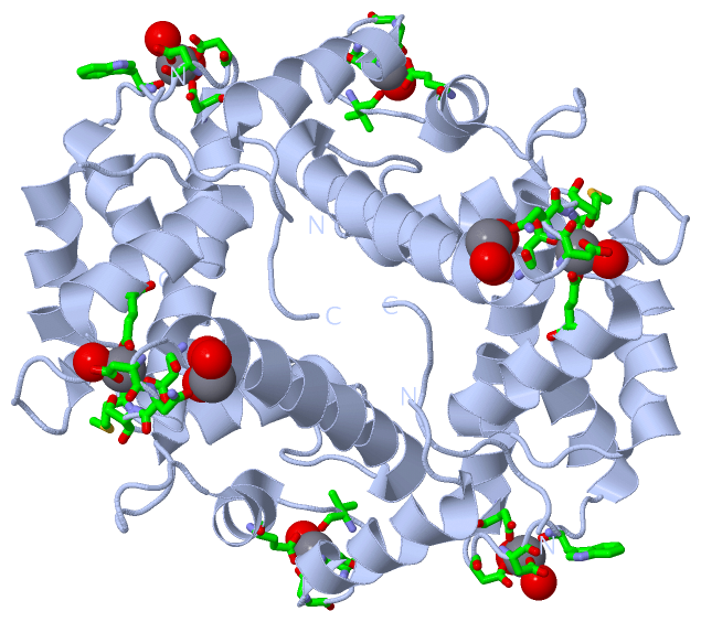
    GO:0005509    calcium ion binding    Interacting selectively and non-covalently with calcium ions (Ca2+).
    GO:0004198    calcium-dependent cysteine-type endopeptidase activity    Catalysis of the hydrolysis of nonterminal peptide bonds in a polypeptide chain by a mechanism using a cysteine residue at the enzyme active center, and requiring the presence of calcium.
    GO:0048306    calcium-dependent protein binding    Interacting selectively and non-covalently with any protein or protein complex (a complex of two or more proteins that may include other nonprotein molecules), in the presence of calcium.
    GO:0042802    identical protein binding    Interacting selectively and non-covalently with an identical protein or proteins.
    GO:0046872    metal ion binding    Interacting selectively and non-covalently with any metal ion.
    GO:0043495    protein anchor    Interacting selectively and non-covalently with both a protein or protein complex and a membrane, in order to maintain the localization of the protein at a specific membrane location.
    GO:0005515    protein binding    Interacting selectively and non-covalently with any protein or protein complex (a complex of two or more proteins that may include other nonprotein molecules).
    GO:0046983    protein dimerization activity    The formation of a protein dimer, a macromolecular structure consists of two noncovalently associated identical or nonidentical subunits.
    GO:0042803    protein homodimerization activity    Interacting selectively and non-covalently with an identical protein to form a homodimer.
biological process
    GO:0006919    activation of cysteine-type endopeptidase activity involved in apoptotic process    Any process that initiates the activity of the inactive enzyme cysteine-type endopeptidase in the context of an apoptotic process.
    GO:0001525    angiogenesis    Blood vessel formation when new vessels emerge from the proliferation of pre-existing blood vessels.
    GO:0006915    apoptotic process    A programmed cell death process which begins when a cell receives an internal (e.g. DNA damage) or external signal (e.g. an extracellular death ligand), and proceeds through a series of biochemical events (signaling pathway phase) which trigger an execution phase. The execution phase is the last step of an apoptotic process, and is typically characterized by rounding-up of the cell, retraction of pseudopodes, reduction of cellular volume (pyknosis), chromatin condensation, nuclear fragmentation (karyorrhexis), plasma membrane blebbing and fragmentation of the cell into apoptotic bodies. When the execution phase is completed, the cell has died.
    GO:0034605    cellular response to heat    Any process that results in a change in state or activity of a cell (in terms of movement, secretion, enzyme production, gene expression, etc.) as a result of a heat stimulus, a temperature stimulus above the optimal temperature for that organism.
    GO:0006886    intracellular protein transport    The directed movement of proteins in a cell, including the movement of proteins between specific compartments or structures within a cell, such as organelles of a eukaryotic cell.
    GO:0032007    negative regulation of TOR signaling    Any process that stops, prevents, or reduces the frequency, rate or extent of TOR signaling.
    GO:0051898    negative regulation of protein kinase B signaling    Any process that stops, prevents, or reduces the frequency, rate or extent of protein kinase B signaling, a series of reactions mediated by the intracellular serine/threonine kinase protein kinase B.
    GO:0030948    negative regulation of vascular endothelial growth factor receptor signaling pathway    Any process that stops, prevents, or reduces the frequency, rate or extent of vascular endothelial growth factor receptor signaling pathway activity.
    GO:0045766    positive regulation of angiogenesis    Any process that activates or increases angiogenesis.
    GO:0043280    positive regulation of cysteine-type endopeptidase activity involved in apoptotic process    Any process that activates or increases the activity of a cysteine-type endopeptidase involved in the apoptotic process.
    GO:0010595    positive regulation of endothelial cell migration    Any process that increases the rate, frequency, or extent of the orderly movement of an endothelial cell into the extracellular matrix to form an endothelium.
    GO:0001938    positive regulation of endothelial cell proliferation    Any process that activates or increases the rate or extent of endothelial cell proliferation.
    GO:0051592    response to calcium ion    Any process that results in a change in state or activity of a cell or an organism (in terms of movement, secretion, enzyme production, gene expression, etc.) as a result of a calcium ion stimulus.
    GO:0036324    vascular endothelial growth factor receptor-2 signaling pathway    Any series of molecular signals initiated by the binding of an extracellular ligand to a vascular endothelial growth factor receptor-2 (VEGFR-2) located on the surface of the receiving cell, and ending with regulation of a downstream cellular process, e.g. transcription.
cellular component
    GO:0005737    cytoplasm    All of the contents of a cell excluding the plasma membrane and nucleus, but including other subcellular structures.
    GO:0031410    cytoplasmic vesicle    A vesicle found in the cytoplasm of a cell.
    GO:0005783    endoplasmic reticulum    The irregular network of unit membranes, visible only by electron microscopy, that occurs in the cytoplasm of many eukaryotic cells. The membranes form a complex meshwork of tubular channels, which are often expanded into slitlike cavities called cisternae. The ER takes two forms, rough (or granular), with ribosomes adhering to the outer surface, and smooth (with no ribosomes attached).
    GO:0070971    endoplasmic reticulum exit site    An endoplasmic reticulum part at which COPII-coated vesicles are produced.
    GO:0005789    endoplasmic reticulum membrane    The lipid bilayer surrounding the endoplasmic reticulum.
    GO:0005768    endosome    A vacuole to which materials ingested by endocytosis are delivered.
    GO:0070062    extracellular exosome    A vesicle that is released into the extracellular region by fusion of the limiting endosomal membrane of a multivesicular body with the plasma membrane. Extracellular exosomes, also simply called exosomes, have a diameter of about 40-100 nm.
    GO:0016020    membrane    A lipid bilayer along with all the proteins and protein complexes embedded in it an attached to it.
    GO:0031965    nuclear membrane    Either of the lipid bilayers that surround the nucleus and form the nuclear envelope; excludes the intermembrane space.
    GO:0005634    nucleus    A membrane-bounded organelle of eukaryotic cells in which chromosomes are housed and replicated. In most cells, the nucleus contains all of the cell's chromosomes except the organellar chromosomes, and is the site of RNA synthesis and processing. In some species, or in specialized cell types, RNA metabolism or DNA replication may be absent.

 Visualization

(-) Interactive Views

Asymmetric Unit
  Complete Structure
    Jena3D(integrated viewing of ligand, site, SAP, PROSITE, SCOP information)
    WebMol | AstexViewer[tm]@PDBe
(Java Applets, require no local installation except for Java; loading may be slow)
    STRAP
(Java WebStart application, automatic local installation, requires Java; full application with system access!)
    RasMol
(require local installation)
    Molscript (VRML)
(requires installation of a VRML viewer; select preferred view via VRML and generate a mono or stereo PDF format file)
 
  Ligands, Modified Residues, Ions
    CA  [ RasMol | Jena3D ]  +environment [ RasMol | Jena3D ]
 
  Sites
    AC1  [ RasMol ]  +environment [ RasMol ]
    AC2  [ RasMol ]  +environment [ RasMol ]
    AC3  [ RasMol ]  +environment [ RasMol ]
    AC4  [ RasMol ]  +environment [ RasMol ]
 
  Cis Peptide Bonds
(no "Cis Peptide Bonds" information available for 1hqv)
 
Biological Units
  Complete Structure
    Biological Unit 1  [ Jena3D ]
    Biological Unit 2  [ Jena3D ]
    Biological Unit 3  [ Jena3D ]

(-) Still Images

Jmol
  protein: cartoon or spacefill or dots and stick; nucleic acid: cartoon and stick; ligands: spacefill; active site: stick
Molscript
  protein, nucleic acid: cartoon; ligands: spacefill; active site: ball and stick

 Databases and Analysis Tools

(-) Databases

Access by PDB/NDB ID
  1hqv
    Family and Domain InformationProDom | SYSTERS
    General Structural InformationGlycoscienceDB | MMDB | NDB | OCA | PDB | PDBe | PDBj | PDBsum | PDBWiki | PQS | PROTEOPEDIA
    Orientation in MembranesOPM
    Protein SurfaceSURFACE
    Secondary StructureDSSP (structure derived) | HSSP (homology derived)
    Structural GenomicsGeneCensus
    Structural NeighboursCE | VAST
    Structure ClassificationCATH | Dali | SCOP
    Validation and Original DataBMRB Data View | BMRB Restraints Grid | EDS | PROCHECK | RECOORD | WHAT_CHECK
 
Access by UniProt ID/Accession number
  PDCD6_MOUSE | P12815
    Comparative Protein Structure ModelsModBase
    Genomic InformationEnsembl
    Protein-protein InteractionDIP
    Sequence, Family and Domain InformationInterPro | Pfam | SMART | UniProtKB/SwissProt
 
Access by Enzyme Classificator   (EC Number)
  (no 'Enzyme Classificator' available)
    General Enzyme InformationBRENDA | EC-PDB | Enzyme | IntEnz
    PathwayKEGG | MetaCyc
 
Access by Disease Identifier   (MIM ID)
  (no 'MIM ID' available)
    Disease InformationOMIM
 
Access by GenAge ID
  (no 'GenAge ID' available)
    Age Related InformationGenAge

(-) Analysis Tools

Access by PDB/NDB ID
    Domain InformationXDom
    Interatomic Contacts of Structural UnitsCSU
    Ligand-protein ContactsLPC
    Protein CavitiescastP
    Sequence and Secondary StructurePDBCartoon
    Structure AlignmentSTRAP(Java WebStart application, automatic local installation, requires Java; full application with system access!)
    Structure and Sequence BrowserSTING
 
Access by UniProt ID/Accession number
  PDCD6_MOUSE | P12815
    Protein Disorder PredictionDisEMBL | FoldIndex | GLOBPLOT (for more information see DisProt)

 Related Entries

(-) Entries Sharing at Least One Protein Chain (UniProt ID)

UniProtKB/Swiss-Prot
        PDCD6_MOUSE | P128155jjg

(-) Related Entries Specified in the PDB File

(no "Related Entries Specified in the PDB File" available for 1HQV)