|
|
|
|
 Description
|
|
Compounds
|
||||||||||||||||||||||||||||||||||||||||||||||||||
Chains, Units
Summary Information (see also Sequences/Alignments below) |
Ligands, Modified Residues, Ions (1, 4)
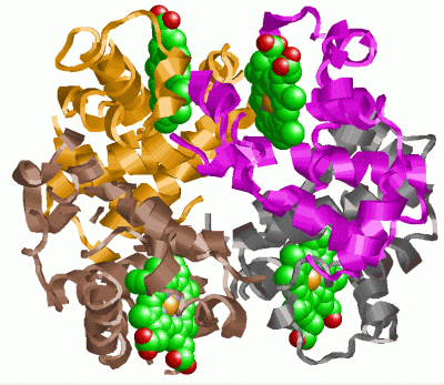
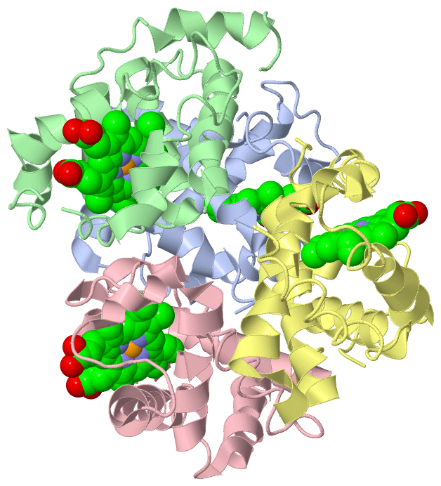
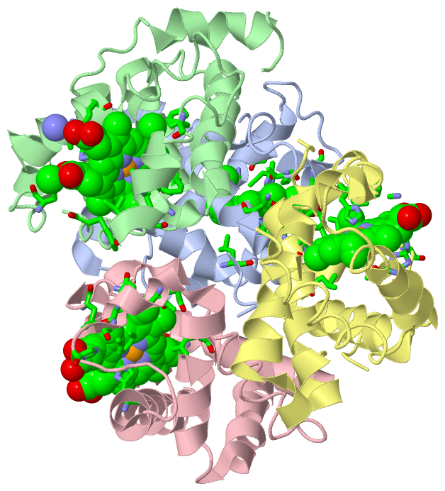
Asymmetric/Biological Unit (1, 4)
|
Sites (4, 4)
Asymmetric Unit (4, 4)
|
SS Bonds (0, 0)| (no "SS Bond" information available for 1HDS) |
Cis Peptide Bonds (17, 17)
Asymmetric/Biological Unit
|
||||||||||||||||||||||||||||||||||||||||||||||||||||||||||||||||||||||||
SAPs(SNPs)/Variants (3, 6)
Asymmetric/Biological Unit (3, 6)
|
||||||||||||||||||||||||||||||||||||||||||||||||||||||||||||||||||||||||||||||||||||
PROSITE Motifs (1, 4)| Asymmetric/Biological Unit (1, 4) |
Exons (0, 0)| (no "Exon" information available for 1HDS) |
Sequences/Alignments
Asymmetric/Biological UnitChain A from PDB Type:PROTEIN Length:141 aligned with HBA_ODOVI | P01972 from UniProtKB/Swiss-Prot Length:141 Alignment length:141 10 20 30 40 50 60 70 80 90 100 110 120 130 140 HBA_ODOVI 1 VLSAABKSBVKAAWGKVGGNAAPYGAZALZRMFLSFPTTKTYFPHFBLSHGSAZVKAHGZKVABALTKAVGHLBBLPGTLSBLSBLHAHKLRVBPVBFKLLSHSLLVTLATHLPBBFTPAVHASLBKFLABVSTVLTSKYR 141 SCOP domains d1hdsa_ A: Hemoglobin, alpha-chain SCOP domains CATH domains 1hdsA00 A:1-141 Globins CATH domains Pfam domains --------------------------------------------------------------------------------------------------------------------------------------------- Pfam domains SAPs(SNPs) ----T--------------K---F--------------------------------------------------------------------------------------------------------------------- SAPs(SNPs) PROSITE -GLOBIN PDB: A:2-141 UniProt: 2-141 PROSITE Transcript --------------------------------------------------------------------------------------------------------------------------------------------- Transcript 1hds A 1 VLSAANKSNVKAAWGKVGGNAPAYGAQALQRMFLSFPTTKTYFPHFDLSHGSAQQKAHGQKVANALTKAQGHLNDLPGTLSNLSNLHAHKLRVNPVNFKLLSHSLLVTLASHLPTNFTPAVHANLNKFLANDSTVLTSKYR 141 10 20 30 40 50 60 70 80 90 100 110 120 130 140 Chain B from PDB Type:PROTEIN Length:145 aligned with HBB_ODOVI | P02074 from UniProtKB/Swiss-Prot Length:145 Alignment length:145 10 20 30 40 50 60 70 80 90 100 110 120 130 140 HBB_ODOVI 1 MLTAEEKAAVTGFWGKVNVDVVGAEALGRLLVVYPWTQRFFEHFGDLSSAGAVMGNPKVKAHGKRVLDAFSEGLKHLDDLKGAFAELSELHCNKLHVDPENFRLLGNVLVVVLARNFGGEFTPLVQADFQKVVAGVANALAHRYH 145 SCOP domains d1hdsb_ B: Hemoglobin, beta-chain SCOP domains CATH domains 1hdsB00 B:1-145 Globins CATH domains Pfam domains ------------------------------------------------------------------------------------------------------------------------------------------------- Pfam domains SAPs(SNPs) ------------------------------------------------------------------------------------------------------------------------------------------------- SAPs(SNPs) PROSITE (2) -GLOBIN PDB: B:2-145 UniProt: 2-145 PROSITE (2) Transcript ------------------------------------------------------------------------------------------------------------------------------------------------- Transcript 1hds B 1 MLTAEEKAAVTGFWGKVDVDVVGAQALGRLLVVYPWTQRFFQHFGNLSSAGAVMNNPKVKAHGKRVLDAFTQGLKHLDDLKGAFAQLSGLHCNKLHVNPQNFRLLGNVLALVVARNFGGQFTPNVQALFQKVVAGVANALAHKYH 145 10 20 30 40 50 60 70 80 90 100 110 120 130 140 Chain C from PDB Type:PROTEIN Length:141 aligned with HBA_ODOVI | P01972 from UniProtKB/Swiss-Prot Length:141 Alignment length:141 10 20 30 40 50 60 70 80 90 100 110 120 130 140 HBA_ODOVI 1 VLSAABKSBVKAAWGKVGGNAAPYGAZALZRMFLSFPTTKTYFPHFBLSHGSAZVKAHGZKVABALTKAVGHLBBLPGTLSBLSBLHAHKLRVBPVBFKLLSHSLLVTLATHLPBBFTPAVHASLBKFLABVSTVLTSKYR 141 SCOP domains d1hdsc_ C: Hemoglobin, alpha-chain SCOP domains CATH domains 1hdsC00 C:1-141 Globins CATH domains Pfam domains --------------------------------------------------------------------------------------------------------------------------------------------- Pfam domains SAPs(SNPs) ----T--------------K---F--------------------------------------------------------------------------------------------------------------------- SAPs(SNPs) PROSITE -GLOBIN PDB: C:2-141 UniProt: 2-141 PROSITE Transcript --------------------------------------------------------------------------------------------------------------------------------------------- Transcript 1hds C 1 VLSAANKSNVKAAWGKVGGNAPAYGAQALQRMFLSFPTTKTYFPHFDLSHGSAQQKAHGQKVANALTKAQGHLNDLPGTLSNLSNLHAHKLRVNPVNFKLLSHSLLVTLASHLPTNFTPAVHANLNKFLANDSTVLTSKYR 141 10 20 30 40 50 60 70 80 90 100 110 120 130 140 Chain D from PDB Type:PROTEIN Length:145 aligned with HBB_ODOVI | P02074 from UniProtKB/Swiss-Prot Length:145 Alignment length:145 10 20 30 40 50 60 70 80 90 100 110 120 130 140 HBB_ODOVI 1 MLTAEEKAAVTGFWGKVNVDVVGAEALGRLLVVYPWTQRFFEHFGDLSSAGAVMGNPKVKAHGKRVLDAFSEGLKHLDDLKGAFAELSELHCNKLHVDPENFRLLGNVLVVVLARNFGGEFTPLVQADFQKVVAGVANALAHRYH 145 SCOP domains d1hdsd_ D: Hemoglobin, beta-chain SCOP domains CATH domains 1hdsD00 D:1-145 Globins CATH domains Pfam domains ------------------------------------------------------------------------------------------------------------------------------------------------- Pfam domains SAPs(SNPs) ------------------------------------------------------------------------------------------------------------------------------------------------- SAPs(SNPs) PROSITE (2) -GLOBIN PDB: D:2-145 UniProt: 2-145 PROSITE (2) Transcript ------------------------------------------------------------------------------------------------------------------------------------------------- Transcript 1hds D 1 MLTAEEKAAVTGFWGKVDVDVVGAQALGRLLVVYPWTQRFFQHFGNLSSAGAVMNNPKVKAHGKRVLDAFTQGLKHLDDLKGAFAQLSGLHCNKLHVNPQNFRLLGNVLALVVARNFGGQFTPNVQALFQKVVAGVANALAHKYH 145 10 20 30 40 50 60 70 80 90 100 110 120 130 140
|
||||||||||||||||||||
SCOP Domains (2, 4)
Asymmetric/Biological Unit
 |
CATH Domains (1, 4)
Asymmetric/Biological Unit
|
Pfam Domains (0, 0)| (no "Pfam Domain" information available for 1HDS) |
Gene Ontology (8, 16)|
Asymmetric/Biological Unit(hide GO term definitions) Chain A,C (HBA_ODOVI | P01972)
Chain B,D (HBB_ODOVI | P02074)
|
||||||||||||||||||||||||||||||||||||||||||||||||||||||||||||||||||||||||||||||||||||||||||||||||||||||||||||||||||||||||||||||||||||
Interactive Views
|
||||||||||||||||||||||||||||||||||||||||||||||||||||||||||||||||||||||||||||||||||||||||||||||||||||||||||||||||||||||||||||||||||||||||||||||||||||||||||||||||||||||||||||||||||||||||||||||||||||||||||||||||||||||||||||||||||||||||||||||||||||||||||||
Still Images
|
||||||||||||||||
Databases
|
||||||||||||||||||||||||||||||||||||||||||||||||||||||||||||||||||||||||||||||||||||||||||||||||||||||||||||||||||||||||||||||||||||||||||||||||||||||||||||||||||||||||||||||||||||||||||
Analysis Tools
|
||||||||||||||||||||||||||||||||||||||||||||||||||||||||||||||||||||||||
Entries Sharing at Least One Protein Chain (UniProt ID)
Related Entries Specified in the PDB File
|
|