| molecular function |
|---|
| | GO:0008745 | | N-acetylmuramoyl-L-alanine amidase activity | | Catalysis of the hydrolysis of the link between N-acetylmuramoyl residues and L-amino acid residues in certain bacterial cell-wall glycopeptides. |
| | GO:0016787 | | hydrolase activity | | Catalysis of the hydrolysis of various bonds, e.g. C-O, C-N, C-C, phosphoric anhydride bonds, etc. Hydrolase is the systematic name for any enzyme of EC class 3. |
| biological process |
|---|
| | GO:0071555 | | cell wall organization | | A process that results in the assembly, arrangement of constituent parts, or disassembly of the cell wall, the rigid or semi-rigid envelope lying outside the cell membrane of plant, fungal and most prokaryotic cells, maintaining their shape and protecting them from osmotic lysis. |
| | GO:0030420 | | establishment of competence for transformation | | The process in which a naturally transformable bacterium acquires the ability to take up exogenous DNA. This term should be applied only to naturally transformable bacteria, and should not be used in the context of artificially induced bacterial transformation. |
| | GO:0009253 | | peptidoglycan catabolic process | | The chemical reactions and pathways resulting in the breakdown of peptidoglycans, any of a class of glycoconjugates found in bacterial cell walls. |
| | GO:0030435 | | sporulation resulting in formation of a cellular spore | | The process in which a relatively unspecialized cell acquires the specialized features of a cellular spore, a cell form that can be used for dissemination, for survival of adverse conditions because of its heat and dessication resistance, and/or for reproduction. |
| cellular component |
|---|
| | GO:0005576 | | extracellular region | | The space external to the outermost structure of a cell. For cells without external protective or external encapsulating structures this refers to space outside of the plasma membrane. This term covers the host cell environment outside an intracellular parasite. |
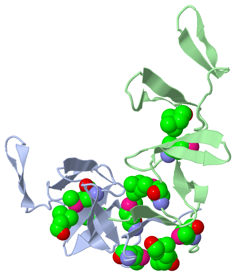
 Description
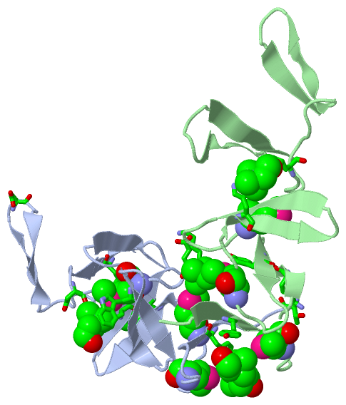