|
|
|
|
 Description
|
|
Compounds
|
||||||||||||||||||||||||
Chains, Units
Summary Information (see also Sequences/Alignments below) |
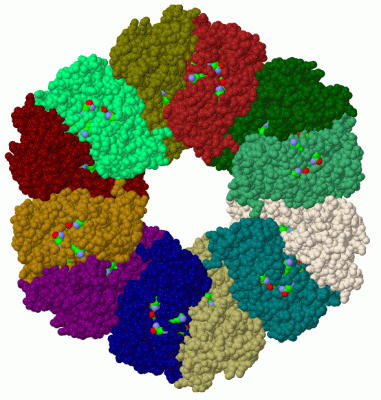
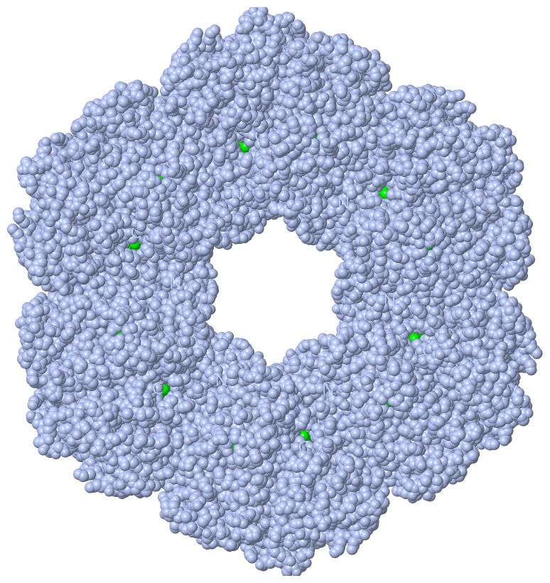
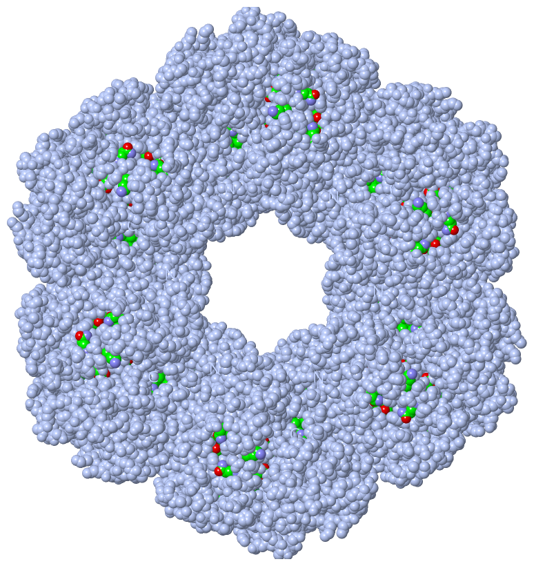
Ligands, Modified Residues, Ions (4, 5)| Asymmetric Unit (4, 5) Biological Unit 1 (2, 24) |
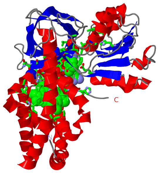
Sites (5, 5)
Asymmetric Unit (5, 5)
|
SS Bonds (0, 0)| (no "SS Bond" information available for 1GVH) |
Cis Peptide Bonds (0, 0)| (no "Cis Peptide Bond" information available for 1GVH) |
SAPs(SNPs)/Variants (0, 0)| (no "SAP(SNP)/Variant" information available for 1GVH) |
PROSITE Motifs (2, 2)| Asymmetric Unit (2, 2) Biological Unit 1 (2, 24) |
Exons (0, 0)| (no "Exon" information available for 1GVH) |
Sequences/Alignments
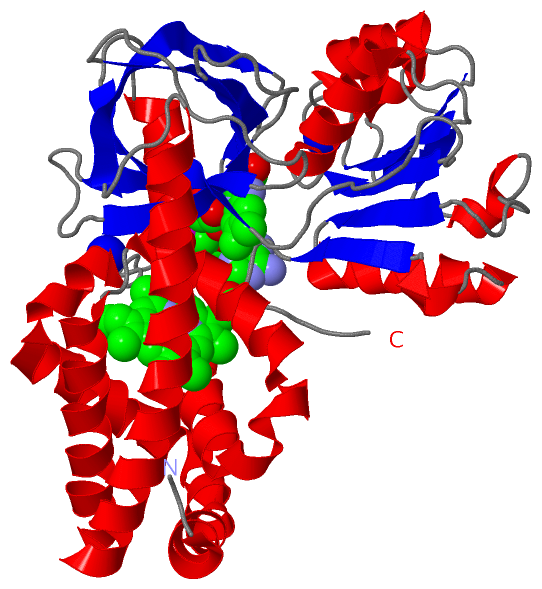
Asymmetric UnitChain A from PDB Type:PROTEIN Length:396 aligned with HMP_ECOLI | P24232 from UniProtKB/Swiss-Prot Length:396 Alignment length:396 10 20 30 40 50 60 70 80 90 100 110 120 130 140 150 160 170 180 190 200 210 220 230 240 250 260 270 280 290 300 310 320 330 340 350 360 370 380 390 HMP_ECOLI 1 MLDAQTIATVKATIPLLVETGPKLTAHFYDRMFTHNPELKEIFNMSNQRNGDQREALFNAIAAYASNIENLPALLPAVEKIAQKHTSFQIKPEQYNIVGEHLLATLDEMFSPGQEVLDAWGKAYGVLANVFINREAEIYNENASKAGGWEGTRDFRIVAKTPRSALITSFELEPVDGGAVAEYRPGQYLGVWLKPEGFPHQEIRQYSLTRKPDGKGYRIAVKREEGGQVSNWLHNHANVGDVVKLVAPAGDFFMAVADDTPVTLISAGVGQTPMLAMLDTLAKAGHTAQVNWFHAAENGDVHAFADEVKELGQSLPRFTAHTWYRQPSEADRAKGQFDSEGLMDLSKLEGAFSDPTMQFYLCGPVGFMQFTAKQLVDLGVKQENIHYECFGPHKVL 396 SCOP domains d1gvha1 A:1-146 Flavohemoglobin, N-terminal domain d1gvha2 A:147-253 Flavohemoglobin, central domain d1gvha3 A:254-396 Flavohemoglobin, C-terminal domain SCOP domains CATH domains 1gvhA01 A:1-148 Globins 1gvhA02 A:149-254 Translation factors 1gvhA03 A:255-396 Nucleotide-binding domain of ferredoxin-NADP reductase (FNR) module CATH domains Pfam domains ------------------------------------------------------------------------------------------------------------------------------------------------------------------------------------------------------------------------------------------------------------------------------------------------------------------------------------------------------------------------------------------------------------ Pfam domains SAPs(SNPs) ------------------------------------------------------------------------------------------------------------------------------------------------------------------------------------------------------------------------------------------------------------------------------------------------------------------------------------------------------------------------------------------------------------ SAPs(SNPs) PROSITE GLOBIN PDB: A:1-134 UniProt: 1-134 ---------------FAD_FR PDB: A:150-255 UniProt: 150-255 --------------------------------------------------------------------------------------------------------------------------------------------- PROSITE Transcript ------------------------------------------------------------------------------------------------------------------------------------------------------------------------------------------------------------------------------------------------------------------------------------------------------------------------------------------------------------------------------------------------------------ Transcript 1gvh A 1 MLDAQTIATVKATIPLLVETGPKLTAHFYDRMFTHNPELKEIFNMSNQRNGDQREALFNAIAAYASNIENLPALLPAVEKIAQKHTSFQIKPEQYNIVGEHLLATLDEMFSPGQEVLDAWGKAYGVLANVFINREAEIYNENASKAGGWEGTRDFRIVAKTPRSALITSFELEPVDGGAVAEYRPGQYLGVWLKPEGFPHQEIRQYSLTRKPDGKGYRIAVKREEGGQVSNWLHNHANVGDVVKLVAPAGDFFMAVADDTPVTLISAGVGQTPMLAMLDTLAKAGHTAQVNWFHAAENGDVHAFADEVKELGQSLPRFTAHTWYRQPSEADRAKGQFDSEGLMDLSKLEGAFSDPTMQFYLCGPVGFMQFTAKQLVDLGVKQENIHYECFGPHKVL 396 10 20 30 40 50 60 70 80 90 100 110 120 130 140 150 160 170 180 190 200 210 220 230 240 250 260 270 280 290 300 310 320 330 340 350 360 370 380 390
|
||||||||||||||||||||
SCOP Domains (3, 3)
Asymmetric Unit

|
CATH Domains (3, 3)
Asymmetric Unit
|
Pfam Domains (0, 0)| (no "Pfam Domain" information available for 1GVH) |
Gene Ontology (15, 15)|
Asymmetric Unit(hide GO term definitions) Chain A (HMP_ECOLI | P24232)
|
||||||||||||||||||||||||||||||||||||||||||||||||||||||||||||||||||||||||||||||||||||||||||||||||||||||||||||
Interactive Views
|
|||||||||||||||||||||||||||||||||||||||||||||||||||||||||||||||||||||||||||||||||||||||||||||||||||||||||||||||||||||||||||||||||||||||||||||||||||||||||||||||||||||||||||||||||||||||||
Still Images
|
||||||||||||||||
Databases
|
||||||||||||||||||||||||||||||||||||||||||||||||||||||||||||||||||||||||||||||||||||||||||||||||||||||||||||||||||||||||||||||||||||||||||||||||||||||||||||||||
Analysis Tools
|
|||||||||||||||||||||||||||||||||||||||||||||||||||||||||||||
Entries Sharing at Least One Protein Chain (UniProt ID)
Related Entries Specified in the PDB File
|
|