|
|
|
|
 Description
|
|
Compounds
|
||||||||||||||||||||||||||||
Chains, Units
Summary Information (see also Sequences/Alignments below) |
Ligands, Modified Residues, Ions (1, 2)
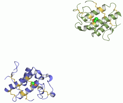
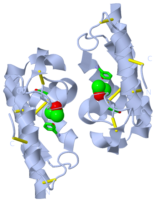
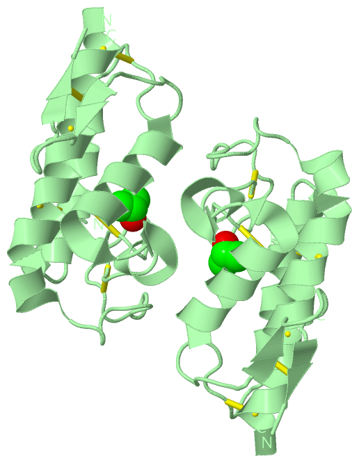
Asymmetric Unit (1, 2)
|
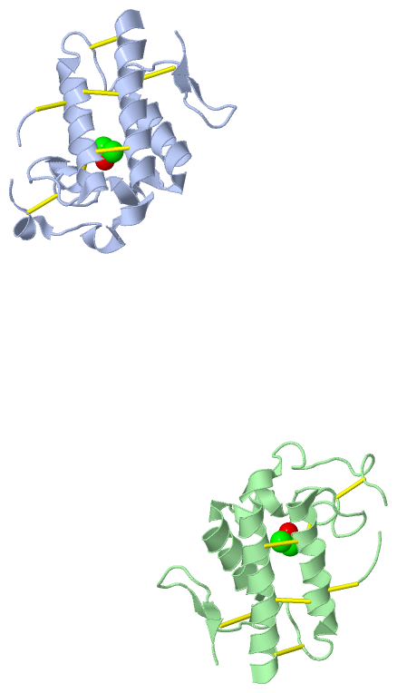
Sites (2, 2)
Asymmetric Unit (2, 2)
|
SS Bonds (14, 14)
Asymmetric Unit
|
||||||||||||||||||||||||||||||||||||||||||||||||||||||||||||
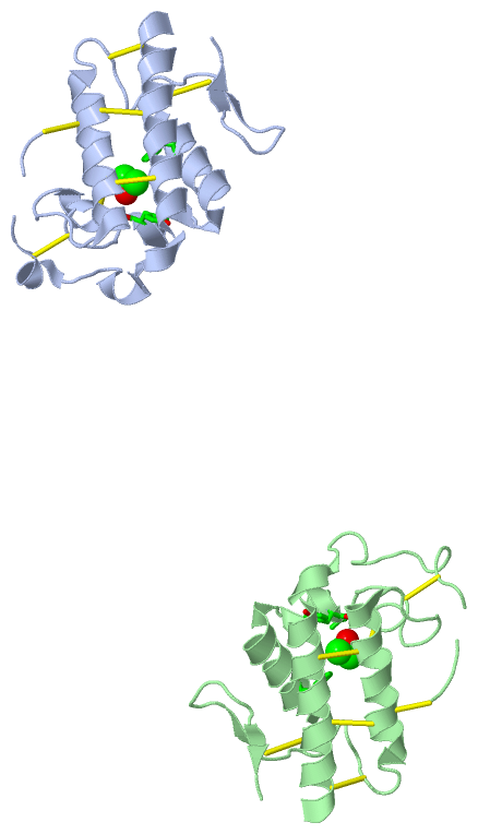
Cis Peptide Bonds (2, 2)
Asymmetric Unit
|
||||||||||||
SAPs(SNPs)/Variants (0, 0)| (no "SAP(SNP)/Variant" information available for 1GMZ) |
PROSITE Motifs (2, 4)
Asymmetric Unit (2, 4)
|
||||||||||||||||||||||||||||||||||||||||||||||||||||||||||||||||||||||||||||||||||||||||||||||||
Exons (0, 0)| (no "Exon" information available for 1GMZ) |
Sequences/Alignments
Asymmetric UnitChain A from PDB Type:PROTEIN Length:119 aligned with PA2B3_BOTPI | P58464 from UniProtKB/Swiss-Prot Length:120 Alignment length:122 82 113 81 | 112 | 10 20 30 40 50 60 70 80| | 89 99 109 | | 118 PA2B3_BOTPI 1 DLWQFGQMILKETGKLPFPYYTYGGCYCGVGGRRGLGTKDDRCCYVHDCCYKKLTGCPKTDDRYSYSWLDLTIVCGEDDPC-KELCECDKAIAVCFRENLGTYNKKYRYHLKP-CKKADKPC 120 SCOP domains d1gmza_ A: Snake phospholipase A2 SCOP domains CATH domains 1gmzA00 A:1-122 Phospholipase A2 CATH domains Pfam domains -------------------------------------------------------------------------------------------------------------------------- Pfam domains SAPs(SNPs) -------------------------------------------------------------------------------------------------------------------------- SAPs(SNPs) PROSITE ------------------------------------------PA2_HIS ----------------------------------PA2_ASP --------------------------- PROSITE Transcript -------------------------------------------------------------------------------------------------------------------------- Transcript 1gmz A 1 DLWQFGKMILKETGKLPFPYYVTYGCYCGVGGRGGPKDATDRCCFVHDCCYGKLTSCKPKTDRYSYSRKDGTIVCGE-DPCRKEICECDKAAAVCFRENLDTYNKKYMSYLKSLCKK--DDC 122 10 20 30 40 50 60 70 | 80 90 100 110 |120 77 | 117 | 79 120 Chain B from PDB Type:PROTEIN Length:120 aligned with PA2B3_BOTPI | P58464 from UniProtKB/Swiss-Prot Length:120 Alignment length:122 82 113 81 | 112 | 10 20 30 40 50 60 70 80| | 89 99 109 | | 118 PA2B3_BOTPI 1 DLWQFGQMILKETGKLPFPYYTYGGCYCGVGGRRGLGTKDDRCCYVHDCCYKKLTGCPKTDDRYSYSWLDLTIVCGEDDPC-KELCECDKAIAVCFRENLGTYNKKYRYHLKP-CKKADKPC 120 SCOP domains d1gmzb_ B: Snake phospholipase A2 SCOP domains CATH domains 1gmzB00 B:1-122 Phospholipase A2 CATH domains Pfam domains -------------------------------------------------------------------------------------------------------------------------- Pfam domains SAPs(SNPs) -------------------------------------------------------------------------------------------------------------------------- SAPs(SNPs) PROSITE ------------------------------------------PA2_HIS ----------------------------------PA2_ASP --------------------------- PROSITE Transcript -------------------------------------------------------------------------------------------------------------------------- Transcript 1gmz B 1 DLWQFGKMILKETGKLPFPYYVTYGCYCGVGGRGGPKDATDRCCFVHDCCYGKLTSCKPKTDRYSYSRKDGTIVCGE-DPCRKEICECDKAAAVCFRENLDTYNKKYMSYLKSLCKK-ADDC 122 10 20 30 40 50 60 70 | 80 90 100 110 |120 77 | 117 | 79 119
|
||||||||||||||||||||
SCOP Domains (1, 2)
Asymmetric Unit
 |
CATH Domains (1, 2)
Asymmetric Unit
|
Pfam Domains (0, 0)| (no "Pfam Domain" information available for 1GMZ) |
Gene Ontology (7, 7)|
Asymmetric Unit(hide GO term definitions) Chain A,B (PA2B3_BOTPI | P58464)
|
||||||||||||||||||||||||||||||||||||||||||||||||||||||||||||
Interactive Views
|
||||||||||||||||||||||||||||||||||||||||||||||||||||||||||||||||||||||||||||||||||||||||||||||||||||||||||||||||||||||||||||||||||||||||||||||||||||||||||||
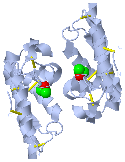
Still Images
|
||||||||||||||||
Databases
|
||||||||||||||||||||||||||||||||||||||||||||||||||||||||||||||||||||||||||||||||||||||||||||||||||||||||||||||||||||||||||||||||||||||||||||||||||||||||||||||||
Analysis Tools
|
|||||||||||||||||||||||||||||||||||||||||||||||||||||||||||||
Entries Sharing at Least One Protein Chain (UniProt ID)
Related Entries Specified in the PDB File
|
|