| molecular function |
|---|
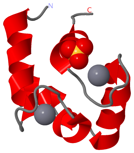
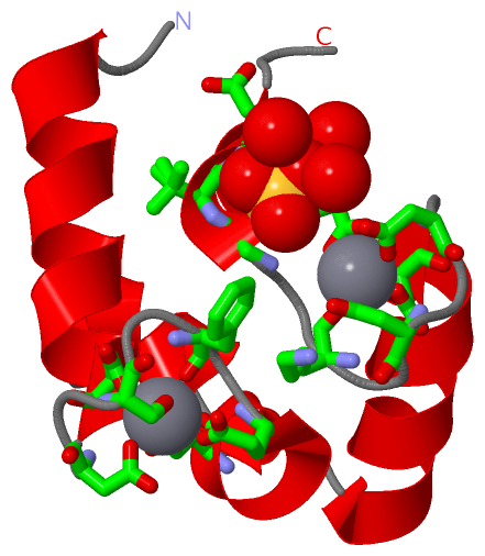
| | GO:0005509 | | calcium ion binding | | Interacting selectively and non-covalently with calcium ions (Ca2+). |
| | GO:0046872 | | metal ion binding | | Interacting selectively and non-covalently with any metal ion. |
| | GO:0046982 | | protein heterodimerization activity | | Interacting selectively and non-covalently with a nonidentical protein to form a heterodimer. |
| | GO:0042803 | | protein homodimerization activity | | Interacting selectively and non-covalently with an identical protein to form a homodimer. |
| biological process |
|---|
| | GO:0051480 | | regulation of cytosolic calcium ion concentration | | Any process involved in the maintenance of an internal steady state of calcium ions within the cytosol of a cell or between the cytosol and its surroundings. |
| cellular component |
|---|
| | GO:0030424 | | axon | | The long process of a neuron that conducts nerve impulses, usually away from the cell body to the terminals and varicosities, which are sites of storage and release of neurotransmitter. |
| | GO:0005737 | | cytoplasm | | All of the contents of a cell excluding the plasma membrane and nucleus, but including other subcellular structures. |
| | GO:0070062 | | extracellular exosome | | A vesicle that is released into the extracellular region by fusion of the limiting endosomal membrane of a multivesicular body with the plasma membrane. Extracellular exosomes, also simply called exosomes, have a diameter of about 40-100 nm. |
| | GO:0043025 | | neuronal cell body | | The portion of a neuron that includes the nucleus, but excludes cell projections such as axons and dendrites. |
| | GO:0005634 | | nucleus | | A membrane-bounded organelle of eukaryotic cells in which chromosomes are housed and replicated. In most cells, the nucleus contains all of the cell's chromosomes except the organellar chromosomes, and is the site of RNA synthesis and processing. In some species, or in specialized cell types, RNA metabolism or DNA replication may be absent. |
| | GO:0043234 | | protein complex | | A stable macromolecular complex composed (only) of two or more polypeptide subunits along with any covalently attached molecules (such as lipid anchors or oligosaccharide) or non-protein prosthetic groups (such as nucleotides or metal ions). Prosthetic group in this context refers to a tightly bound cofactor. The component polypeptide subunits may be identical. |
| | GO:0043195 | | terminal bouton | | Terminal inflated portion of the axon, containing the specialized apparatus necessary to release neurotransmitters. The axon terminus is considered to be the whole region of thickening and the terminal bouton is a specialized region of it. |
 Description