Show PDB file:   
         Plain Text   HTML   (compressed file size)
QuickSearch:   
by PDB,NDB,UniProt,PROSITE Code or Search Term(s)  
(-)Asym./Biol. Unit - manually
(-)Asym./Biol. Unit
(-)Asym./Biol. Unit - sites
collapse expand < >
Image Asym./Biol. Unit - manually
Asym./Biol. Unit - manually  (Jmol Viewer)
Image Asym./Biol. Unit
Asym./Biol. Unit  (Jmol Viewer)
Image Asym./Biol. Unit - sites
Asym./Biol. Unit - sites  (Jmol Viewer)

(-) Description

Title :  SPECIFIC MUTATIONS IN KRAIT PLA2 LEAD TO DIMERIZATION OF PROTEIN MOLECULES: CRYSTAL STRUCTURE OF KRAIT PLA2 AT 2.1 RESOLUTION
 
Authors :  T. P. Singh, S. Gourinath, S. Sharma, G. Singh
Date :  10 Oct 00  (Deposition) - 17 Jun 03  (Release) - 24 Feb 09  (Revision)
Method :  X-RAY DIFFRACTION
Resolution :  2.18
Chains :  Asym./Biol. Unit :  A,B
Keywords :  Structure, Phospholipase A2, Homodimer, Bungarus Caeruleus, Toxin (Keyword Search: [Gene Ontology, PubMed, Web (Google))
 
Reference :  S. Gourinath, G. Singh, S. Sharma, S. Bhanumathi, C. Betzel, M. Paramsivam, A. Srinivasan, T. P. Singh
Specific Mutations In Krait Pla2 Lead To Dimerization Of Protein Molecules: Crystal Structure Of Krait Pla2 At 2. 1 Resolution.
To Be Published
PubMed: search
(for further references see the PDB file header)

(-) Compounds

Molecule 1 - PHOSPHOLIPASE A2
    ChainsA, B
    EC Number3.1.1.4
    FragmentRESIDUES 28-145
    Organism ScientificBUNGARUS CAERULEUS
    Organism Taxid132961
    Other DetailsNATURAL PROTEIN-ISOFORM
    SecretionVENOM

 Structural Features

(-) Chains, Units

  12
Asymmetric/Biological Unit AB

Summary Information (see also Sequences/Alignments below)

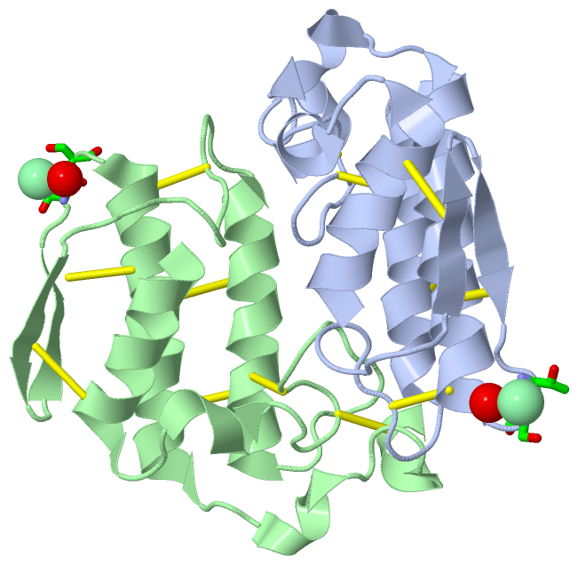
(-) Ligands, Modified Residues, Ions  (1, 2)

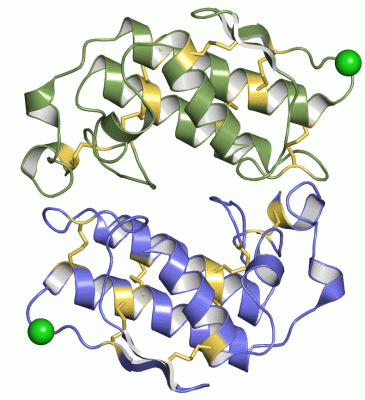
Asymmetric/Biological Unit (1, 2)
No.NameCountTypeFull Name
1CL2Ligand/IonCHLORIDE ION

(-) Sites  (2, 2)

Asymmetric Unit (2, 2)
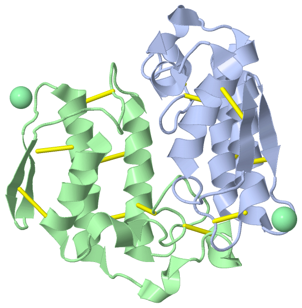
No.NameEvidenceResiduesDescription
1AC1SOFTWARETHR B:82 , SER B:83 , HOH B:186BINDING SITE FOR RESIDUE CL B 121
2AC2SOFTWARETHR A:82 , SER A:83 , HOH A:167BINDING SITE FOR RESIDUE CL A 121

(-) SS Bonds  (14, 14)

Asymmetric/Biological Unit
No.Residues
1A:11 -A:72
2A:27 -A:119
3A:29 -A:45
4A:44 -A:100
5A:51 -A:93
6A:61 -A:86
7A:79 -A:91
8B:11 -B:72
9B:27 -B:119
10B:29 -B:45
11B:44 -B:100
12B:51 -B:93
13B:61 -B:86
14B:79 -B:91

(-) Cis Peptide Bonds  (2, 2)

Asymmetric/Biological Unit
No.Residues
1Lys A:74 -Pro A:75
2Lys B:74 -Pro B:75

 Sequence-Structure Mapping

(-) SAPs(SNPs)/Variants  (0, 0)

(no "SAP(SNP)/Variant" information available for 1G0Z)

(-) PROSITE Motifs  (2, 4)

Asymmetric/Biological Unit (2, 4)
 PROSITEUniProtKBPDB
No.IDACDescriptionIDLocationCountLocation
1PA2_HISPS00118 Phospholipase A2 histidine active site.PA2B5_BUNCE61-68
 
  2A:44-51
B:44-51
2PA2_ASPPS00119 Phospholipase A2 aspartic acid active site.PA2B5_BUNCE107-117
 
  2A:90-100
B:90-100

(-) Exons   (0, 0)

(no "Exon" information available for 1G0Z)

(-) Sequences/Alignments

Asymmetric/Biological Unit
   Reformat: Number of residues per line =  ('0' or empty: single-line sequence representation)
  Number of residues per labelling interval =   
  UniProt sequence: complete  aligned part    
   Show mapping: SCOP domains CATH domains Pfam domains Secondary structure (by author)
SAPs(SNPs) PROSITE motifs Exons
(details for a mapped element are shown in a popup box when the mouse pointer rests over it)
Chain A from PDB  Type:PROTEIN  Length:118
 aligned with PA2B5_BUNCE | Q6SLM1 from UniProtKB/Swiss-Prot  Length:137

    Alignment length:118
                                    29        39        49        59        69        79        89        99       109       119       129        
          PA2B5_BUNCE    20 NLKQFKNMIQCAGTRTWTSYIGYGCYCGYGGSGTPVDELDRCCYTHDHCYNKAANIPGCNPLIKTYSYTCTKPNITCNDTSDSCARFICDCDRTAAICFASAPYNINNIMISASTSCQ 137
               SCOP domains d1g0za_ A: Snake phospholipase A2                                                                                      SCOP domains
               CATH domains 1g0zA00 A:1-120 Phospholipase A2                                                                                       CATH domains
               Pfam domains ---------------------------------------------------------------------------------------------------------------------- Pfam domains
         Sec.struct. author hhhhhhhhhhhhh..hhhhhh...............hhhhhhhhhhhhhhhhhh.............eeee..eeee....hhhhhhhhhhhhhhhhhhhhh..hhhhh....hhhhh Sec.struct. author
                 SAPs(SNPs) ---------------------------------------------------------------------------------------------------------------------- SAPs(SNPs)
                    PROSITE -----------------------------------------PA2_HIS --------------------------------------PA2_ASP    -------------------- PROSITE
                 Transcript ---------------------------------------------------------------------------------------------------------------------- Transcript
                 1g0z A   1 NLKQFKNMIQCAGTRTWTSYIGYGCYCGYGGSGTPVDELDRCCYTHDHCYNKAANIPGCNPLIKTYSYTCTKPNITCNDTSDSCARFICDCDRTAAICFASAPYNINNIMISASTSCQ 120
                                    10   ||   22        32        42        52        62        72        82        92       102       112        
                                        14|                                                                                                       
                                         17                                                                                                       

Chain B from PDB  Type:PROTEIN  Length:118
 aligned with PA2B5_BUNCE | Q6SLM1 from UniProtKB/Swiss-Prot  Length:137

    Alignment length:118
                                    29        39        49        59        69        79        89        99       109       119       129        
          PA2B5_BUNCE    20 NLKQFKNMIQCAGTRTWTSYIGYGCYCGYGGSGTPVDELDRCCYTHDHCYNKAANIPGCNPLIKTYSYTCTKPNITCNDTSDSCARFICDCDRTAAICFASAPYNINNIMISASTSCQ 137
               SCOP domains d1g0zb_ B: Snake phospholipase A2                                                                                      SCOP domains
               CATH domains 1g0zB00 B:1-120 Phospholipase A2                                                                                       CATH domains
               Pfam domains ---------------------------------------------------------------------------------------------------------------------- Pfam domains
         Sec.struct. author hhhhhhhhhhhhh..hhhhh................hhhhhhhhhhhhhhhhhh.............eeee..eeee....hhhhhhhhhhhhhhhhhhhhh..hhhhh....hhhhh Sec.struct. author
                 SAPs(SNPs) ---------------------------------------------------------------------------------------------------------------------- SAPs(SNPs)
                    PROSITE -----------------------------------------PA2_HIS --------------------------------------PA2_ASP    -------------------- PROSITE
                 Transcript ---------------------------------------------------------------------------------------------------------------------- Transcript
                 1g0z B   1 NLKQFKNMIQCAGTRTWTSYIGYGCYCGYGGSGTPVDELDRCCYTHDHCYNKAANIPGCNPLIKTYSYTCTKPNITCNDTSDSCARFICDCDRTAAICFASAPYNINNIMISASTSCQ 120
                                    10   ||   22        32        42        52        62        72        82        92       102       112        
                                        14|                                                                                                       
                                         17                                                                                                       

   Legend:   → Mismatch (orange background)
  - → Gap (green background, '-', border residues have a numbering label)
    → Modified Residue (blue background, lower-case, 'x' indicates undefined single-letter code, labelled with number + name)
  x → Chemical Group (purple background, 'x', labelled with number + name, e.g. ACE or NH2)
  extra numbering lines below/above indicate numbering irregularities and modified residue names etc., number ends below/above '|'

 Classification and Annotation

(-) SCOP Domains  (1, 2)

Asymmetric/Biological Unit

(-) CATH Domains  (1, 2)

Asymmetric/Biological Unit

(-) Pfam Domains  (0, 0)

(no "Pfam Domain" information available for 1G0Z)

(-) Gene Ontology  (6, 6)

Asymmetric/Biological Unit(hide GO term definitions)
Chain A,B   (PA2B5_BUNCE | Q6SLM1)
molecular function
    GO:0005509    calcium ion binding    Interacting selectively and non-covalently with calcium ions (Ca2+).
    GO:0016787    hydrolase activity    Catalysis of the hydrolysis of various bonds, e.g. C-O, C-N, C-C, phosphoric anhydride bonds, etc. Hydrolase is the systematic name for any enzyme of EC class 3.
    GO:0046872    metal ion binding    Interacting selectively and non-covalently with any metal ion.
    GO:0004623    phospholipase A2 activity    Catalysis of the reaction: phosphatidylcholine + H2O = 1-acylglycerophosphocholine + a carboxylate.
biological process
    GO:0016042    lipid catabolic process    The chemical reactions and pathways resulting in the breakdown of lipids, compounds soluble in an organic solvent but not, or sparingly, in an aqueous solvent.
cellular component
    GO:0005576    extracellular region    The space external to the outermost structure of a cell. For cells without external protective or external encapsulating structures this refers to space outside of the plasma membrane. This term covers the host cell environment outside an intracellular parasite.

 Visualization

(-) Interactive Views

Asymmetric/Biological Unit
  Complete Structure
    Jena3D(integrated viewing of ligand, site, SAP, PROSITE, SCOP information)
    WebMol | AstexViewer[tm]@PDBe
(Java Applets, require no local installation except for Java; loading may be slow)
    STRAP
(Java WebStart application, automatic local installation, requires Java; full application with system access!)
    RasMol
(require local installation)
    Molscript (VRML)
(requires installation of a VRML viewer; select preferred view via VRML and generate a mono or stereo PDF format file)
 
  Ligands, Modified Residues, Ions
    CL  [ RasMol | Jena3D ]  +environment [ RasMol | Jena3D ]
 
  Sites
    AC1  [ RasMol ]  +environment [ RasMol ]
    AC2  [ RasMol ]  +environment [ RasMol ]
 
  Cis Peptide Bonds
    Lys A:74 - Pro A:75   [ RasMol ]  
    Lys B:74 - Pro B:75   [ RasMol ]  
 

(-) Still Images

Jmol
  protein: cartoon or spacefill or dots and stick; nucleic acid: cartoon and stick; ligands: spacefill; active site: stick
Molscript
  protein, nucleic acid: cartoon; ligands: spacefill; active site: ball and stick

 Databases and Analysis Tools

(-) Databases

Access by PDB/NDB ID
  1g0z
    Family and Domain InformationProDom | SYSTERS
    General Structural InformationGlycoscienceDB | MMDB | NDB | OCA | PDB | PDBe | PDBj | PDBsum | PDBWiki | PQS | PROTEOPEDIA
    Orientation in MembranesOPM
    Protein SurfaceSURFACE
    Secondary StructureDSSP (structure derived) | HSSP (homology derived)
    Structural GenomicsGeneCensus
    Structural NeighboursCE | VAST
    Structure ClassificationCATH | Dali | SCOP
    Validation and Original DataBMRB Data View | BMRB Restraints Grid | EDS | PROCHECK | RECOORD | WHAT_CHECK
 
Access by UniProt ID/Accession number
  PA2B5_BUNCE | Q6SLM1
    Comparative Protein Structure ModelsModBase
    Genomic InformationEnsembl
    Protein-protein InteractionDIP
    Sequence, Family and Domain InformationInterPro | Pfam | SMART | UniProtKB/SwissProt
 
Access by Enzyme Classificator   (EC Number)
  3.1.1.4
    General Enzyme InformationBRENDA | EC-PDB | Enzyme | IntEnz
    PathwayKEGG | MetaCyc
 
Access by Disease Identifier   (MIM ID)
  (no 'MIM ID' available)
    Disease InformationOMIM
 
Access by GenAge ID
  (no 'GenAge ID' available)
    Age Related InformationGenAge

(-) Analysis Tools

Access by PDB/NDB ID
    Domain InformationXDom
    Interatomic Contacts of Structural UnitsCSU
    Ligand-protein ContactsLPC
    Protein CavitiescastP
    Sequence and Secondary StructurePDBCartoon
    Structure AlignmentSTRAP(Java WebStart application, automatic local installation, requires Java; full application with system access!)
    Structure and Sequence BrowserSTING
 
Access by UniProt ID/Accession number
  PA2B5_BUNCE | Q6SLM1
    Protein Disorder PredictionDisEMBL | FoldIndex | GLOBPLOT (for more information see DisProt)

 Related Entries

(-) Entries Sharing at Least One Protein Chain (UniProt ID)

UniProtKB/Swiss-Prot
        PA2B5_BUNCE | Q6SLM11u4j

(-) Related Entries Specified in the PDB File

1fe5 THREE DIMENSIONAL STRUCTURE OF A NOVEL PHOSPHOLIPASE A2 FROM INDIAN COMMON KRAIT AT 2.45 RESOLUTION