|
|
|
|
 Description
|
|
Compounds
|
||||||||||||||||||||||||||||||||||||||||||||||||
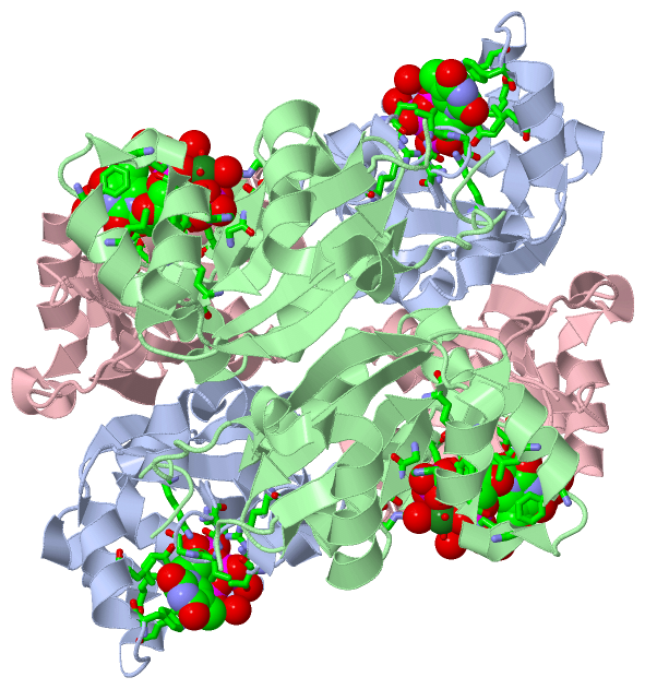
Chains, Units
Summary Information (see also Sequences/Alignments below) |
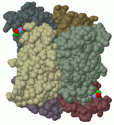
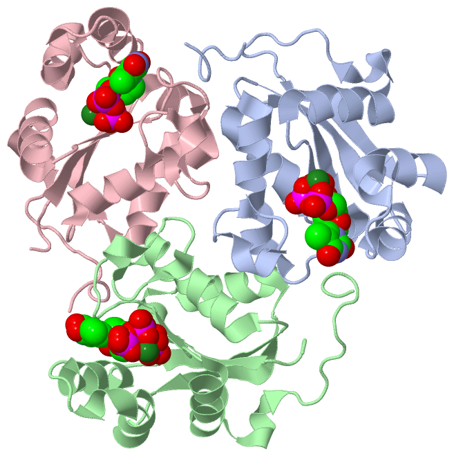
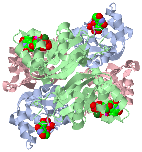
Ligands, Modified Residues, Ions (4, 8)| Asymmetric Unit (4, 8) Biological Unit 1 (3, 10) |
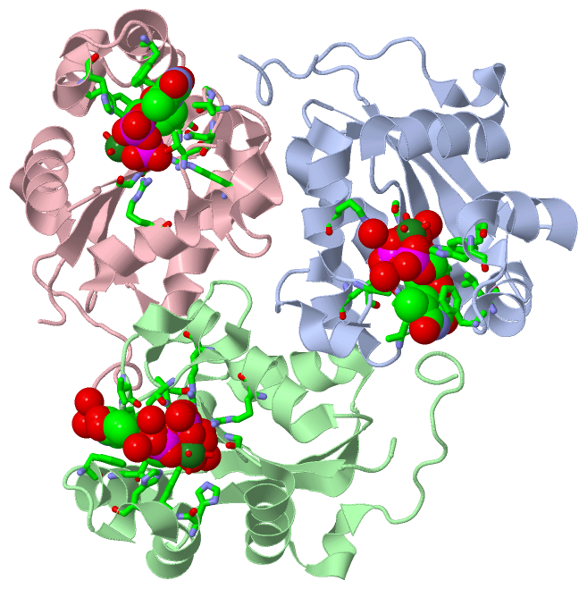
Sites (8, 8)
Asymmetric Unit (8, 8)
|
SS Bonds (0, 0)| (no "SS Bond" information available for 1F3F) |
Cis Peptide Bonds (0, 0)| (no "Cis Peptide Bond" information available for 1F3F) |
SAPs(SNPs)/Variants (0, 0)| (no "SAP(SNP)/Variant" information available for 1F3F) |
PROSITE Motifs (1, 3)
Asymmetric Unit (1, 3)
|
||||||||||||||||||||||||||||||||||||||||||||||||
Exons (0, 0)| (no "Exon" information available for 1F3F) |
Sequences/Alignments
Asymmetric UnitChain A from PDB Type:PROTEIN Length:150 aligned with NDKC_DICDI | P22887 from UniProtKB/Swiss-Prot Length:155 Alignment length:150 15 25 35 45 55 65 75 85 95 105 115 125 135 145 155 NDKC_DICDI 6 VNKERTFLAVKPDGVARGLVGEIIARYEKKGFVLVGLKQLVPTKDLAESHYAEHKERPFFGGLVSFITSGPVVAMVFEGKGVVASARLMIGVTNPLASAPGSIRGDFGVDVGRNIIHGSDSVESANREIALWFKPEELLTEVKPNPNLYE 155 SCOP domains d1f3fa_ A: Nucleoside diphosphate kinase, NDK SCOP domains CATH domains 1f3fA00 A:6-155 [code=3.30.70.141, no name defined] CATH domains Pfam domains ------------------------------------------------------------------------------------------------------------------------------------------------------ Pfam domains SAPs(SNPs) ------------------------------------------------------------------------------------------------------------------------------------------------------ SAPs(SNPs) PROSITE -----------------------------------------------------------------------------------------------------------------NDP_KINAS---------------------------- PROSITE Transcript ------------------------------------------------------------------------------------------------------------------------------------------------------ Transcript 1f3f A 6 VNKERTFLAVKPDGVARGLVGEIIARYEKKGFVLVGLKQLVPTKDLAESHYAEHKERPFFGGLVSFITSGPVVAMVFEGKGVVASARLMIGVTNPLASAPGSIRGDFGVDVGRNIIGGSDSVESANREIALWFKPEELLTEVKPNPNLYE 155 15 25 35 45 55 65 75 85 95 105 115 125 135 145 155 Chain B from PDB Type:PROTEIN Length:150 aligned with NDKC_DICDI | P22887 from UniProtKB/Swiss-Prot Length:155 Alignment length:150 15 25 35 45 55 65 75 85 95 105 115 125 135 145 155 NDKC_DICDI 6 VNKERTFLAVKPDGVARGLVGEIIARYEKKGFVLVGLKQLVPTKDLAESHYAEHKERPFFGGLVSFITSGPVVAMVFEGKGVVASARLMIGVTNPLASAPGSIRGDFGVDVGRNIIHGSDSVESANREIALWFKPEELLTEVKPNPNLYE 155 SCOP domains d1f3fb_ B: Nucleoside diphosphate kinase, NDK SCOP domains CATH domains 1f3fB00 B:6-155 [code=3.30.70.141, no name defined] CATH domains Pfam domains ------------------------------------------------------------------------------------------------------------------------------------------------------ Pfam domains SAPs(SNPs) ------------------------------------------------------------------------------------------------------------------------------------------------------ SAPs(SNPs) PROSITE -----------------------------------------------------------------------------------------------------------------NDP_KINAS---------------------------- PROSITE Transcript ------------------------------------------------------------------------------------------------------------------------------------------------------ Transcript 1f3f B 6 VNKERTFLAVKPDGVARGLVGEIIARYEKKGFVLVGLKQLVPTKDLAESHYAEHKERPFFGGLVSFITSGPVVAMVFEGKGVVASARLMIGVTNPLASAPGSIRGDFGVDVGRNIIGGSDSVESANREIALWFKPEELLTEVKPNPNLYE 155 15 25 35 45 55 65 75 85 95 105 115 125 135 145 155 Chain C from PDB Type:PROTEIN Length:150 aligned with NDKC_DICDI | P22887 from UniProtKB/Swiss-Prot Length:155 Alignment length:150 15 25 35 45 55 65 75 85 95 105 115 125 135 145 155 NDKC_DICDI 6 VNKERTFLAVKPDGVARGLVGEIIARYEKKGFVLVGLKQLVPTKDLAESHYAEHKERPFFGGLVSFITSGPVVAMVFEGKGVVASARLMIGVTNPLASAPGSIRGDFGVDVGRNIIHGSDSVESANREIALWFKPEELLTEVKPNPNLYE 155 SCOP domains d1f3fc_ C: Nucleoside diphosphate kinase, NDK SCOP domains CATH domains 1f3fC00 C:6-155 [code=3.30.70.141, no name defined] CATH domains Pfam domains ------------------------------------------------------------------------------------------------------------------------------------------------------ Pfam domains SAPs(SNPs) ------------------------------------------------------------------------------------------------------------------------------------------------------ SAPs(SNPs) PROSITE -----------------------------------------------------------------------------------------------------------------NDP_KINAS---------------------------- PROSITE Transcript ------------------------------------------------------------------------------------------------------------------------------------------------------ Transcript 1f3f C 6 VNKERTFLAVKPDGVARGLVGEIIARYEKKGFVLVGLKQLVPTKDLAESHYAEHKERPFFGGLVSFITSGPVVAMVFEGKGVVASARLMIGVTNPLASAPGSIRGDFGVDVGRNIIGGSDSVESANREIALWFKPEELLTEVKPNPNLYE 155 15 25 35 45 55 65 75 85 95 105 115 125 135 145 155
|
||||||||||||||||||||
SCOP Domains (1, 3)
Asymmetric Unit
 |
CATH Domains (1, 3)
Asymmetric Unit
|
Pfam Domains (0, 0)| (no "Pfam Domain" information available for 1F3F) |
Gene Ontology (32, 32)|
Asymmetric Unit(hide GO term definitions) Chain A,B,C (NDKC_DICDI | P22887)
|
||||||||||||||||||||||||||||||||||||||||||||||||||||||||||||||||||||||||||||||||||||||||||||||||||||||||||||||||||||||||||||||||||||||||||||||||||||||||||||||||||||||||||||||||||||||||||||||||||||||||||||||||||
Interactive Views
|
||||||||||||||||||||||||||||||||||||||||||||||||||||||||||||||||||||||||||||||||||||||||||||||||||||||||||||||||||||||||||||||||||||||||||||||||||||||||||||||||||||||||||||||||||||||||||||||||||||||||||||||
Still Images
|
||||||||||||||||
Databases
|
||||||||||||||||||||||||||||||||||||||||||||||||||||||||||||||||||||||||||||||||||||||||||||||||||||||||||||||||||||||||||||||||||||||||||||||||||||||||||||||||
Analysis Tools
|
|||||||||||||||||||||||||||||||||||||||||||||||||||||||||||||
Entries Sharing at Least One Protein Chain (UniProt ID)
Related Entries Specified in the PDB File
|
|