Show PDB file:   
         Plain Text   HTML   (compressed file size)
QuickSearch:   
by PDB,NDB,UniProt,PROSITE Code or Search Term(s)  
(-)Asymmetric Unit
(-)Asym. Unit - sites
(-)Biological Unit 1
(-)Biol. Unit 1 - sites
(-)Biological Unit 2
(-)Biological Unit 3
collapse expand < >
Image Asymmetric Unit
Asymmetric Unit  (Jmol Viewer)
Image Asym. Unit - sites
Asym. Unit - sites  (Jmol Viewer)
Image Biological Unit 1
Biological Unit 1  (Jmol Viewer)
Image Biol. Unit 1 - sites
Biol. Unit 1 - sites  (Jmol Viewer)
Image Biological Unit 2
Biological Unit 2  (Jmol Viewer)
Image Biological Unit 3
Biological Unit 3  (Jmol Viewer)

(-) Description

Title :  CRYSTAL STRUCTURE OF MURINE OLFACTORY MARKER PROTEIN
 
Authors :  P. C. Smith, J. F. Hunt, Northeast Structural Genomics Consortium
Date :  31 May 00  (Deposition) - 15 Aug 01  (Release) - 13 Jul 11  (Revision)
Method :  X-RAY DIFFRACTION
Resolution :  2.30
Chains :  Asym. Unit :  A,B
Biol. Unit 1:  A  (1x)
Biol. Unit 2:  B  (1x)
Biol. Unit 3:  A (1x),B (1x)
Keywords :  Beta, Structural Genomics, Psi, Protein Structure Initiative, Northeast Structural Genomics Consortium, Nesg, Signaling Protein (Keyword Search: [Gene Ontology, PubMed, Web (Google))
 
Reference :  P. C. Smith, S. Firestein, J. F. Hunt
The Crystal Structure Of The Olfactory Marker Protein At 2. A Resolution.
J. Mol. Biol. V. 319 807 2002
PubMed-ID: 12054872  |  Reference-DOI: 10.1016/S0022-2836(02)00242-5

(-) Compounds

Molecule 1 - OLFACTORY MARKER PROTEIN
    ChainsA, B
    EngineeredYES
    Expression SystemESCHERICHIA COLI
    Expression System PlasmidPET28A
    Expression System Taxid562
    MutationYES
    Organism CommonMOUSE
    Organism ScientificMUS MUSCULUS
    Organism Taxid10090
    Other DetailsCDNA LIBRARY

 Structural Features

(-) Chains, Units

  12
Asymmetric Unit AB
Biological Unit 1 (1x)A 
Biological Unit 2 (1x) B
Biological Unit 3 (1x)A (1x)B (1x)

Summary Information (see also Sequences/Alignments below)

(-) Ligands, Modified Residues, Ions  (3, 21)

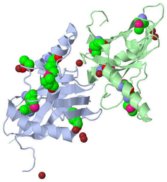
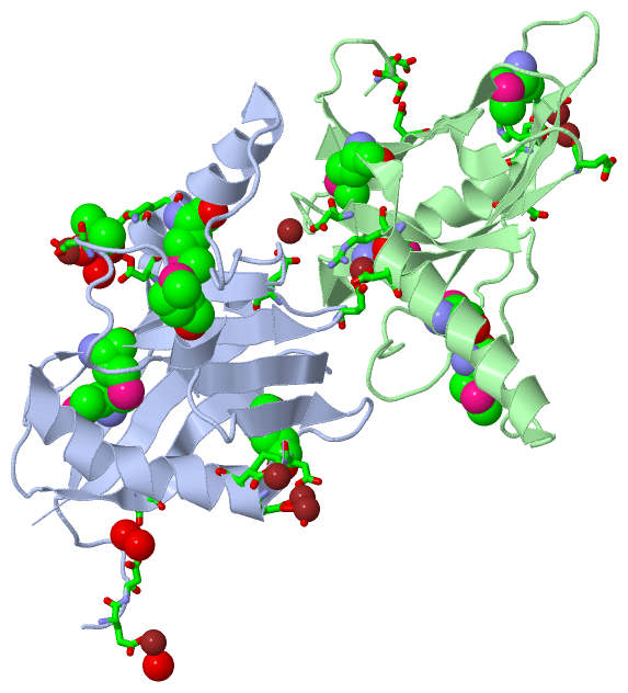
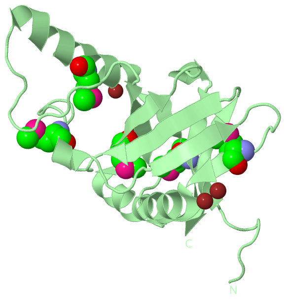
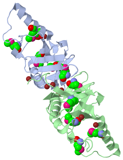
Asymmetric Unit (3, 21)
No.NameCountTypeFull Name
1CAC1Ligand/IonCACODYLATE ION
2MSE10Mod. Amino AcidSELENOMETHIONINE
3ZN10Ligand/IonZINC ION
Biological Unit 1 (2, 6)
No.NameCountTypeFull Name
1CAC1Ligand/IonCACODYLATE ION
2MSE5Mod. Amino AcidSELENOMETHIONINE
3ZN-1Ligand/IonZINC ION
Biological Unit 2 (1, 5)
No.NameCountTypeFull Name
1CAC-1Ligand/IonCACODYLATE ION
2MSE5Mod. Amino AcidSELENOMETHIONINE
3ZN-1Ligand/IonZINC ION
Biological Unit 3 (2, 6)
No.NameCountTypeFull Name
1CAC1Ligand/IonCACODYLATE ION
2MSE5Mod. Amino AcidSELENOMETHIONINE
3ZN-1Ligand/IonZINC ION

(-) Sites  (11, 11)

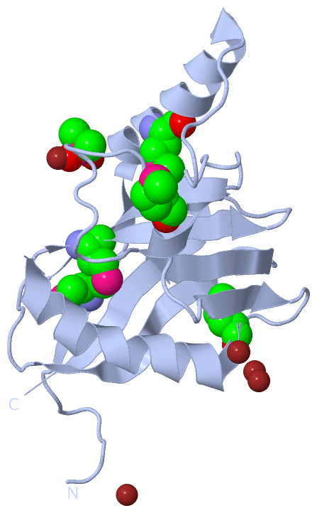
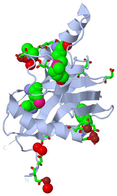
Asymmetric Unit (11, 11)
No.NameEvidenceResiduesDescription
01AC1SOFTWAREASP A:116 , TRP A:117 , GLU A:119 , ZN A:302 , ASP B:1066BINDING SITE FOR RESIDUE ZN A 301
02AC2SOFTWAREASP A:90 , CAC A:351 , HOH A:556 , GLU B:1147BINDING SITE FOR RESIDUE ZN A 304
03AC3SOFTWAREASP A:121 , HOH A:502 , HOH A:561 , GLU B:1003BINDING SITE FOR RESIDUE ZN A 305
04AC4SOFTWAREASP A:18 , ASP A:20 , CAC A:351 , HOH A:557BINDING SITE FOR RESIDUE ZN A 306
05AC5SOFTWAREGLU A:3 , HOH A:610 , HOH A:646 , ASP B:1121BINDING SITE FOR RESIDUE ZN A 307
06AC6SOFTWAREASP A:66 , ZN B:309 , ASP B:1116 , TRP B:1117 , GLU B:1119BINDING SITE FOR RESIDUE ZN B 308
07AC7SOFTWAREASP A:4 , ASP A:66 , HOH A:541 , HOH A:614 , ZN B:308 , GLU B:1119BINDING SITE FOR RESIDUE ZN B 309
08AC8SOFTWAREGLU A:150 , ASP B:1056BINDING SITE FOR RESIDUE ZN B 310
09AC9SOFTWAREASP A:18 , ASP A:20 , GLN A:24 , ASP A:90 , ZN A:304 , ZN A:306 , HOH A:629 , GLU B:1147BINDING SITE FOR RESIDUE CAC A 351
10BC1SOFTWAREGLU A:119 , ZN A:301 , HOH A:677 , ASP B:1004 , ASP B:1066BINDING SITE FOR RESIDUE ZN A 302
11BC2SOFTWAREGLU A:147 , HOH B:678 , ASP B:1018 , ASP B:1020 , ARG B:1054BINDING SITE FOR RESIDUE ZN B 303

(-) SS Bonds  (0, 0)

(no "SS Bond" information available for 1F35)

(-) Cis Peptide Bonds  (2, 2)

Asymmetric Unit
No.Residues
1Asp A:101 -Pro A:102
2Asp B:1101 -Pro B:1102

 Sequence-Structure Mapping

(-) SAPs(SNPs)/Variants  (0, 0)

(no "SAP(SNP)/Variant" information available for 1F35)

(-) PROSITE Motifs  (0, 0)

(no "PROSITE Motif" information available for 1F35)

(-) Exons   (0, 0)

(no "Exon" information available for 1F35)

(-) Sequences/Alignments

Asymmetric Unit
   Reformat: Number of residues per line =  ('0' or empty: single-line sequence representation)
  Number of residues per labelling interval =   
  UniProt sequence: complete  aligned part    
   Show mapping: SCOP domains CATH domains Pfam domains Secondary structure (by author)
SAPs(SNPs) PROSITE motifs Exons
(details for a mapped element are shown in a popup box when the mouse pointer rests over it)
Chain A from PDB  Type:PROTEIN  Length:162
 aligned with OMP_MOUSE | Q64288 from UniProtKB/Swiss-Prot  Length:163

    Alignment length:162
                                    11        21        31        41        51        61        71        81        91       101       111       121       131       141       151       161  
           OMP_MOUSE      2 AEDGPQKQQLEMPLVLDQDLTQQMRLRVESLKQRGEKKQDGEKLIRPAESVYRLDFIQQQKLQFDHWNVVLDKPGKVTITGTSQNWTPDLTNLMTRQLLDPAAIFWRKEDSDAMDWNEADALEFGERLSDLAKIRKVMYFLITFGEGVEPANLKASVVFNQL  163
               SCOP domains d1f35a_ A: Olfactory marker protein                                                                                                                                SCOP domains
               CATH domains 1f35A00 A:2-163  [code=2.60.120.390, no name defined]                                                                                                              CATH domains
               Pfam domains ------------------------------------------------------------------------------------------------------------------------------------------------------------------ Pfam domains
         Sec.struct. author ........eeeee.eehhhhhhhhhhhhhhhhhh..............eeeeeee......eeeeeeeeee...eeeeeee.............hhh.....eeeee......eeehhhhhhhhhhhhhhhhhhh.eeeeeeee....hhh.eeeeeeeee. Sec.struct. author
                 SAPs(SNPs) ------------------------------------------------------------------------------------------------------------------------------------------------------------------ SAPs(SNPs)
                    PROSITE ------------------------------------------------------------------------------------------------------------------------------------------------------------------ PROSITE
                 Transcript ------------------------------------------------------------------------------------------------------------------------------------------------------------------ Transcript
                1f35 A    2 AEDGPQKQQLEmPLVLDQDLTQQmRLRVESLKQRGEKKQDGEKLIRPAESVYRLDFIQQQKLQFDHWNVVLDKPGKVTITGTSQNWTPDLTNLmTRQLLDPAAIFWRKEDSDAmDWNEADALEFGERLSDLAKIRKVmYFLITFGEGVEPANLKASVVFNQL  163
                                    11 |      21   |    31        41        51        61        71        81        91   |   101       111   |   121       131       141       151       161  
                                      13-MSE      25-MSE                                                                95-MSE             115-MSE                 139-MSE                    

Chain B from PDB  Type:PROTEIN  Length:162
 aligned with OMP_MOUSE | Q64288 from UniProtKB/Swiss-Prot  Length:163

    Alignment length:162
                                    11        21        31        41        51        61        71        81        91       101       111       121       131       141       151       161  
           OMP_MOUSE      2 AEDGPQKQQLEMPLVLDQDLTQQMRLRVESLKQRGEKKQDGEKLIRPAESVYRLDFIQQQKLQFDHWNVVLDKPGKVTITGTSQNWTPDLTNLMTRQLLDPAAIFWRKEDSDAMDWNEADALEFGERLSDLAKIRKVMYFLITFGEGVEPANLKASVVFNQL  163
               SCOP domains d1f35b_ B: Olfactory marker protein                                                                                                                                SCOP domains
               CATH domains 1f35B00 B:1002-1163  [code=2.60.120.390, no name defined]                                                                                                          CATH domains
               Pfam domains ------------------------------------------------------------------------------------------------------------------------------------------------------------------ Pfam domains
         Sec.struct. author ........eeeeeeeehhhhhhhhhhhhhhhhhh..............eeeeeee......eeeeeeeeee...eeeeeee.............hhh.....eeeee......eeehhhhhhhhhhhhhhhhhhh.eeeeeeee....hhh.eeeeeeeee. Sec.struct. author
                 SAPs(SNPs) ------------------------------------------------------------------------------------------------------------------------------------------------------------------ SAPs(SNPs)
                    PROSITE ------------------------------------------------------------------------------------------------------------------------------------------------------------------ PROSITE
                 Transcript ------------------------------------------------------------------------------------------------------------------------------------------------------------------ Transcript
                1f35 B 1002 AEDGPQKQQLEmPLVLDQDLTQQmRLRVESLKQRGEKKQDGEKLIRPAESVYRLDFIQQQKLQFDHWNVVLDKPGKVTITGTSQNWTPDLTNLmTRQLLDPAAIFWRKEDSDAmDWNEADALEFGERLSDLAKIRKVmYFLITFGEGVEPANLKASVVFNQL 1163
                                  1011 |    1021   |  1031      1041      1051      1061      1071      1081      1091   |  1101      1111   |  1121      1131      1141      1151      1161  
                                    1013-MSE    1025-MSE                                                              1095-MSE            1115-MSE                1139-MSE                    

   Legend:   → Mismatch (orange background)
  - → Gap (green background, '-', border residues have a numbering label)
    → Modified Residue (blue background, lower-case, 'x' indicates undefined single-letter code, labelled with number + name)
  x → Chemical Group (purple background, 'x', labelled with number + name, e.g. ACE or NH2)
  extra numbering lines below/above indicate numbering irregularities and modified residue names etc., number ends below/above '|'

 Classification and Annotation

(-) SCOP Domains  (1, 2)

Asymmetric Unit

(-) CATH Domains  (1, 2)

Asymmetric Unit
(-)
Class: Mainly Beta (13760)

(-) Pfam Domains  (0, 0)

(no "Pfam Domain" information available for 1F35)

(-) Gene Ontology  (11, 11)

Asymmetric Unit(hide GO term definitions)
Chain A,B   (OMP_MOUSE | Q64288)
molecular function
    GO:0004871    signal transducer activity    Conveys a signal across a cell to trigger a change in cell function or state. A signal is a physical entity or change in state that is used to transfer information in order to trigger a response.
biological process
    GO:0007268    chemical synaptic transmission    The vesicular release of classical neurotransmitter molecules from a presynapse, across a chemical synapse, the subsequent activation of neurotransmitter receptors at the postsynapse of a target cell (neuron, muscle, or secretory cell) and the effects of this activation on the postsynaptic membrane potential and ionic composition of the postsynaptic cytosol. This process encompasses both spontaneous and evoked release of neurotransmitter and all parts of synaptic vesicle exocytosis. Evoked transmission starts with the arrival of an action potential at the presynapse.
    GO:0022008    neurogenesis    Generation of cells within the nervous system.
    GO:0050896    response to stimulus    Any process that results in a change in state or activity of a cell or an organism (in terms of movement, secretion, enzyme production, gene expression, etc.) as a result of a stimulus. The process begins with detection of the stimulus and ends with a change in state or activity or the cell or organism.
    GO:0007608    sensory perception of smell    The series of events required for an organism to receive an olfactory stimulus, convert it to a molecular signal, and recognize and characterize the signal. Olfaction involves the detection of chemical composition of an organism's ambient medium by chemoreceptors. This is a neurological process.
    GO:0007165    signal transduction    The cellular process in which a signal is conveyed to trigger a change in the activity or state of a cell. Signal transduction begins with reception of a signal (e.g. a ligand binding to a receptor or receptor activation by a stimulus such as light), or for signal transduction in the absence of ligand, signal-withdrawal or the activity of a constitutively active receptor. Signal transduction ends with regulation of a downstream cellular process, e.g. regulation of transcription or regulation of a metabolic process. Signal transduction covers signaling from receptors located on the surface of the cell and signaling via molecules located within the cell. For signaling between cells, signal transduction is restricted to events at and within the receiving cell.
cellular component
    GO:0030424    axon    The long process of a neuron that conducts nerve impulses, usually away from the cell body to the terminals and varicosities, which are sites of storage and release of neurotransmitter.
    GO:0005737    cytoplasm    All of the contents of a cell excluding the plasma membrane and nucleus, but including other subcellular structures.
    GO:0005829    cytosol    The part of the cytoplasm that does not contain organelles but which does contain other particulate matter, such as protein complexes.
    GO:0043025    neuronal cell body    The portion of a neuron that includes the nucleus, but excludes cell projections such as axons and dendrites.
    GO:0005634    nucleus    A membrane-bounded organelle of eukaryotic cells in which chromosomes are housed and replicated. In most cells, the nucleus contains all of the cell's chromosomes except the organellar chromosomes, and is the site of RNA synthesis and processing. In some species, or in specialized cell types, RNA metabolism or DNA replication may be absent.

 Visualization

(-) Interactive Views

Asymmetric Unit
  Complete Structure
    Jena3D(integrated viewing of ligand, site, SAP, PROSITE, SCOP information)
    WebMol | AstexViewer[tm]@PDBe
(Java Applets, require no local installation except for Java; loading may be slow)
    STRAP
(Java WebStart application, automatic local installation, requires Java; full application with system access!)
    RasMol
(require local installation)
    Molscript (VRML)
(requires installation of a VRML viewer; select preferred view via VRML and generate a mono or stereo PDF format file)
 
  Ligands, Modified Residues, Ions
    CAC  [ RasMol | Jena3D ]  +environment [ RasMol | Jena3D ]
    MSE  [ RasMol | Jena3D ]  +environment [ RasMol | Jena3D ]
    ZN  [ RasMol | Jena3D ]  +environment [ RasMol | Jena3D ]
 
  Sites
    AC1  [ RasMol ]  +environment [ RasMol ]
    AC2  [ RasMol ]  +environment [ RasMol ]
    AC3  [ RasMol ]  +environment [ RasMol ]
    AC4  [ RasMol ]  +environment [ RasMol ]
    AC5  [ RasMol ]  +environment [ RasMol ]
    AC6  [ RasMol ]  +environment [ RasMol ]
    AC7  [ RasMol ]  +environment [ RasMol ]
    AC8  [ RasMol ]  +environment [ RasMol ]
    AC9  [ RasMol ]  +environment [ RasMol ]
    BC1  [ RasMol ]  +environment [ RasMol ]
    BC2  [ RasMol ]  +environment [ RasMol ]
 
  Cis Peptide Bonds
    Asp A:101 - Pro A:102   [ RasMol ]  
    Asp B:1101 - Pro B:1102   [ RasMol ]  
 
Biological Units
  Complete Structure
    Biological Unit 1  [ Jena3D ]
    Biological Unit 2  [ Jena3D ]
    Biological Unit 3  [ Jena3D ]

(-) Still Images

Jmol
  protein: cartoon or spacefill or dots and stick; nucleic acid: cartoon and stick; ligands: spacefill; active site: stick
Molscript
  protein, nucleic acid: cartoon; ligands: spacefill; active site: ball and stick

 Databases and Analysis Tools

(-) Databases

Access by PDB/NDB ID
  1f35
    Family and Domain InformationProDom | SYSTERS
    General Structural InformationGlycoscienceDB | MMDB | NDB | OCA | PDB | PDBe | PDBj | PDBsum | PDBWiki | PQS | PROTEOPEDIA
    Orientation in MembranesOPM
    Protein SurfaceSURFACE
    Secondary StructureDSSP (structure derived) | HSSP (homology derived)
    Structural GenomicsGeneCensus
    Structural NeighboursCE | VAST
    Structure ClassificationCATH | Dali | SCOP
    Validation and Original DataBMRB Data View | BMRB Restraints Grid | EDS | PROCHECK | RECOORD | WHAT_CHECK
 
Access by UniProt ID/Accession number
  OMP_MOUSE | Q64288
    Comparative Protein Structure ModelsModBase
    Genomic InformationEnsembl
    Protein-protein InteractionDIP
    Sequence, Family and Domain InformationInterPro | Pfam | SMART | UniProtKB/SwissProt
 
Access by Enzyme Classificator   (EC Number)
  (no 'Enzyme Classificator' available)
    General Enzyme InformationBRENDA | EC-PDB | Enzyme | IntEnz
    PathwayKEGG | MetaCyc
 
Access by Disease Identifier   (MIM ID)
  (no 'MIM ID' available)
    Disease InformationOMIM
 
Access by GenAge ID
  (no 'GenAge ID' available)
    Age Related InformationGenAge

(-) Analysis Tools

Access by PDB/NDB ID
    Domain InformationXDom
    Interatomic Contacts of Structural UnitsCSU
    Ligand-protein ContactsLPC
    Protein CavitiescastP
    Sequence and Secondary StructurePDBCartoon
    Structure AlignmentSTRAP(Java WebStart application, automatic local installation, requires Java; full application with system access!)
    Structure and Sequence BrowserSTING
 
Access by UniProt ID/Accession number
  OMP_MOUSE | Q64288
    Protein Disorder PredictionDisEMBL | FoldIndex | GLOBPLOT (for more information see DisProt)

 Related Entries

(-) Entries Sharing at Least One Protein Chain (UniProt ID)

UniProtKB/Swiss-Prot
        OMP_MOUSE | Q642881job 1jod

(-) Related Entries Specified in the PDB File

(no "Related Entries Specified in the PDB File" available for 1F35)