Asymmetric Unit (5, 5)
| No. | Name | Evidence | Residues | Description |
|---|
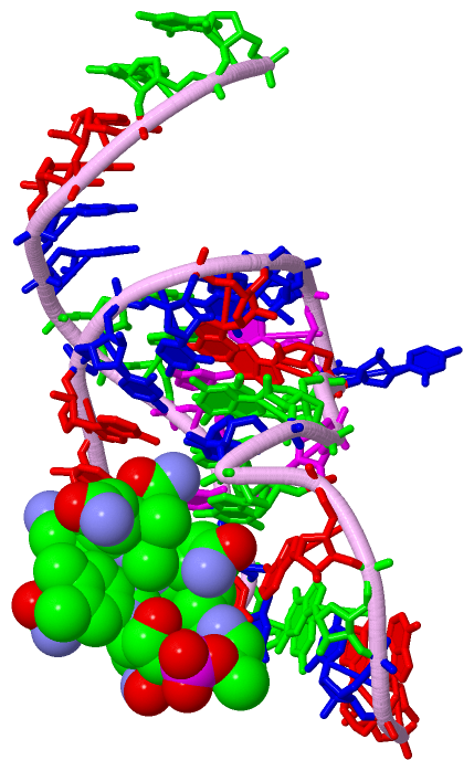
| 1 | AC1 | SOFTWARE | C A:211 , A A:214 , A A:216 , C A:229 , G A:230 , A A:231 , HOH A:1151 , HOH A:1180 , HOH A:1246 , HOH A:1253 , HOH A:1272 , HOH A:1387 , HOH A:1459 , HOH A:1503 , HOH A:1508 , HOH A:1673 , HOH A:1764 , HOH B:1490 , HOH B:1612 , A D:516 , HOH D:1189 , HOH D:1443 | BINDING SITE FOR RESIDUE CNC A 701 |
| 2 | AC2 | SOFTWARE | HOH A:1290 , C B:311 , A B:314 , A B:316 , C B:329 , G B:330 , HOH B:1121 , HOH B:1137 , HOH B:1266 , HOH B:1293 , HOH B:1364 , HOH B:1420 , HOH B:1468 , HOH B:1612 , HOH B:1707 , HOH B:1709 , HOH B:1725 , HOH B:1777 , HOH B:1802 , CNC D:1001 , HOH D:1256 , HOH D:1328 | BINDING SITE FOR RESIDUE CNC B 801 |
| 3 | AC3 | SOFTWARE | C C:411 , A C:414 , A C:416 , C C:429 , G C:430 , HOH C:1109 , HOH C:1139 , HOH C:1178 , HOH C:1211 , HOH C:1236 , HOH C:1258 , HOH C:1271 , HOH C:1454 , A E:117 , HOH E:1314 | BINDING SITE FOR RESIDUE CNC C 901 |
| 4 | AC4 | SOFTWARE | A A:216 , A A:217 , HOH A:1246 , CNC B:801 , HOH B:1725 , C D:511 , A D:514 , A D:516 , C D:529 , G D:530 , HOH D:1107 , HOH D:1143 , HOH D:1168 , HOH D:1232 , HOH D:1255 , HOH D:1256 , HOH D:1288 , HOH D:1328 , HOH D:1332 , HOH D:1409 , HOH D:1591 | BINDING SITE FOR RESIDUE CNC D 1001 |
| 5 | AC5 | SOFTWARE | A C:417 , HOH C:1211 , C E:111 , A E:114 , A E:116 , C E:129 , G E:130 , HOH E:1281 , HOH E:1321 , HOH E:1585 , HOH E:1754 , HOH E:1759 , HOH E:1778 | BINDING SITE FOR RESIDUE CNC E 601 |
|
 Description