|
|
|
|
 Description
|
|
Compounds
|
||||||||||||||||||||||||||||||||||||
Chains, Units
Summary Information (see also Sequences/Alignments below) |
Ligands, Modified Residues, Ions (1, 1)
Asymmetric/Biological Unit (1, 1)
|
Sites (1, 1)
Asymmetric Unit (1, 1)
|
SS Bonds (0, 0)| (no "SS Bond" information available for 1EFD) |
Cis Peptide Bonds (0, 0)| (no "Cis Peptide Bond" information available for 1EFD) |
SAPs(SNPs)/Variants (0, 0)| (no "SAP(SNP)/Variant" information available for 1EFD) |
PROSITE Motifs (1, 1)
Asymmetric/Biological Unit (1, 1)
|
||||||||||||||||||||||||
Exons (0, 0)| (no "Exon" information available for 1EFD) |
Sequences/Alignments
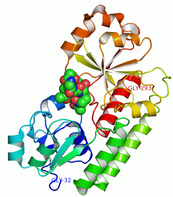
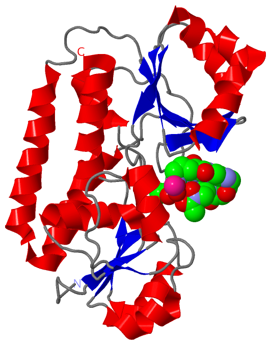
Asymmetric/Biological UnitChain N from PDB Type:PROTEIN Length:262 aligned with FHUD_ECOLI | P07822 from UniProtKB/Swiss-Prot Length:296 Alignment length:262 41 51 61 71 81 91 101 111 121 131 141 151 161 171 181 191 201 211 221 231 241 251 261 271 281 291 FHUD_ECOLI 32 AIDPNRIVALEWLPVELLLALGIVPYGVADTINYRLWVSEPPLPDSVIDVGLRTEPNLELLTEMKPSFMVWSAGYGPSPEMLARIAPGRGFNFSDGKQPLAMARKSLTEMADLLNLQSAAETHLAQYEDFIRSMKPRFVKRGARPLLLTTLIDPRHMLVFGPNSLFQEILDEYGIPNAWQGETNFWGSTAVSIDRLAAYKDVDVLCFDHDNSKDMDALMATPLWQAMPFVRAGRFQRVPAVWFYGATLSAMHFVRVLDNAIG 293 SCOP domains d1efdn_ N: Periplasmic ferric siderophore binding protein FhuD SCOP domains CATH domains 1efdN01 N:32-122 Nitrogenase molybdenum iron protein domain 1efdN02 N:123-293 Nitrogenase molybdenum iron protein domain CATH domains Pfam domains ---------------------------------------------------------------------------------------------------------------------------------------------------------------------------------------------------------------------------------------------------------------------- Pfam domains SAPs(SNPs) ---------------------------------------------------------------------------------------------------------------------------------------------------------------------------------------------------------------------------------------------------------------------- SAPs(SNPs) PROSITE -----FE_B12_PBP PDB: N:37-293 UniProt: 37-296 PROSITE Transcript ---------------------------------------------------------------------------------------------------------------------------------------------------------------------------------------------------------------------------------------------------------------------- Transcript 1efd N 32 GIDPNRIVALEWLPVELLLALGIVPYGVADTINYRLWVSEPPLPDSVIDVGLRTEPNLELLTEMKPSFMVWSAGYGPSPEMLARIAPGRGFNFSDGKQPLAMARKSLTEMADLLNLQSAAETHLAQYEDFIRSMKPRFVKRGARPLLLTTLIDPRHMLVFGPNSLFQEILDEYGIPNAWQGETNFWGSTAVSIDRLAAYKDVDVLCFDHDNSKDMDALMATPLWQAMPFVRAGRFQRVPAVWFYGATLSAMHFVRVLDNAIG 293 41 51 61 71 81 91 101 111 121 131 141 151 161 171 181 191 201 211 221 231 241 251 261 271 281 291
|
||||||||||||||||||||
SCOP Domains (1, 1)
Asymmetric/Biological Unit
 |
CATH Domains (1, 2)
Asymmetric/Biological Unit
|
Pfam Domains (0, 0)| (no "Pfam Domain" information available for 1EFD) |
Gene Ontology (4, 4)|
Asymmetric/Biological Unit(hide GO term definitions) Chain N (FHUD_ECOLI | P07822)
|
||||||||||||||||||||||||||||||||||||
Interactive Views
|
||||||||||||||||||||||||||||||||||||||||||||||||||||||||||||||||||||||||||||||||||||||||||||||||||||||||||||||||||||||
Still Images
|
||||||||||||||||
Databases
|
||||||||||||||||||||||||||||||||||||||||||||||||||||||||||||||||||||||||||||||||||||||||||||||||||||||||||||||||||||||||||||||||||||||||||||||||||||||||||||||||
Analysis Tools
|
|||||||||||||||||||||||||||||||||||||||||||||||||||||||||||||
Entries Sharing at Least One Protein Chain (UniProt ID)
Related Entries Specified in the PDB File
|
|