|
|
|
|
 Description
|
|
Compounds
|
||||||||||||||||||||||||||||||||||||||||||||
Chains, Units
Summary Information (see also Sequences/Alignments below) |
Ligands, Modified Residues, Ions (0, 0)| (no "Ligand,Modified Residues,Ions" information available for 1EF5) |
Sites (0, 0)| (no "Site" information available for 1EF5) |
SS Bonds (0, 0)| (no "SS Bond" information available for 1EF5) |
Cis Peptide Bonds (0, 0)| (no "Cis Peptide Bond" information available for 1EF5) |
SAPs(SNPs)/Variants (0, 0)| (no "SAP(SNP)/Variant" information available for 1EF5) |
PROSITE Motifs (1, 1)
NMR Structure (1, 1)
|
||||||||||||||||||||||||
Exons (0, 0)| (no "Exon" information available for 1EF5) |
Sequences/Alignments
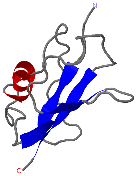
NMR StructureChain A from PDB Type:PROTEIN Length:88 aligned with RGL1_MOUSE | Q60695 from UniProtKB/Swiss-Prot Length:768 Alignment length:88 656 666 676 686 696 706 716 726 RGL1_MOUSE 647 EDTCIIRISVEDNNGNMYKSIMLTSQDKTPAVIQRAMSKHNLESDPAEEYELVQVISEDKELVIPDSANVFYAMNSQVNFDFILRKKN 734 SCOP domains d1ef5a_ A: Rgl SCOP domains CATH domains 1ef5A00 A:647-734 Phosphatidylinositol 3-kinase Catalytic Subunit; Chain A, domain 1 CATH domains Pfam domains ---------------------------------------------------------------------------------------- Pfam domains SAPs(SNPs) ---------------------------------------------------------------------------------------- SAPs(SNPs) PROSITE -RA PDB: A:648-734 UniProt: 648-735 PROSITE Transcript ---------------------------------------------------------------------------------------- Transcript 1ef5 A 647 EDTCIIRISVEDNNGNMYKSIMLTSQDKTPAVIQRAMSKHNLESDPAEEYELVQVISEDKELVIPDSANVFYAMNSQVNFDFILRKKN 734 656 666 676 686 696 706 716 726
|
||||||||||||||||||||
SCOP Domains (1, 1)
NMR Structure
 |
CATH Domains (1, 1)
NMR Structure
|
Pfam Domains (0, 0)| (no "Pfam Domain" information available for 1EF5) |
Gene Ontology (6, 6)|
NMR Structure(hide GO term definitions) Chain A (RGL1_MOUSE | Q60695)
|
||||||||||||||||||||||||||||||||||||||||||||||||||||||
Interactive Views
|
||||||||||||||||||||||||||||||||||||||||||||||||||||||||||||||||||||||||||||||||||||||||||||||||||||||||||||||||||||
Still Images
|
||||||||||||||||
Databases
|
||||||||||||||||||||||||||||||||||||||||||||||||||||||||||||||||||||||||||||||||||||||||||||||||||||||||||||||||||||||||||||||||||||||||||||||||||||||||||||||||
Analysis Tools
|
|||||||||||||||||||||||||||||||||||||||||||||||||||||||||||||
Entries Sharing at Least One Protein Chain (UniProt ID)
Related Entries Specified in the PDB File
|
|