|
|
|
|
 Description
|
|
Compounds
|
||||||||||||||||||||
Chains, Units
Summary Information (see also Sequences/Alignments below) |
Ligands, Modified Residues, Ions (2, 4)| Asymmetric/Biological Unit (2, 4) |
Sites (4, 4)
Asymmetric Unit (4, 4)
|
SS Bonds (0, 0)| (no "SS Bond" information available for 1E29) |
Cis Peptide Bonds (1, 1)
Asymmetric/Biological Unit
|
||||||||
SAPs(SNPs)/Variants (0, 0)| (no "SAP(SNP)/Variant" information available for 1E29) |
PROSITE Motifs (1, 1)
Asymmetric/Biological Unit (1, 1)
|
||||||||||||||||||||||||
Exons (0, 0)| (no "Exon" information available for 1E29) |
Sequences/Alignments
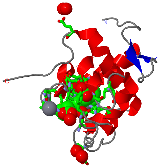
Asymmetric/Biological UnitChain A from PDB Type:PROTEIN Length:135 aligned with CY550_SYNY3 | Q55013 from UniProtKB/Swiss-Prot Length:160 Alignment length:135 35 45 55 65 75 85 95 105 115 125 135 145 155 CY550_SYNY3 26 VELTESTRTIPLDEAGGTTTLTARQFTNGQKIFVDTCTQCHLQGKTKTNNNVSLGLADLAGAEPRRDNVLALVEFLKNPKSYDGEDDYSELHPNISRPDIYPEMRNYTEDDIFDVAGYTLIAPKLDERWGGTIYF 160 SCOP domains d1e29a_ A: Photosystem II associated cytochrome c549 SCOP domains CATH domains 1e29A00 A:1-135 Cytochrome c CATH domains Pfam domains --------------------------------------------------------------------------------------------------------------------------------------- Pfam domains SAPs(SNPs) --------------------------------------------------------------------------------------------------------------------------------------- SAPs(SNPs) PROSITE -----------------------CYTC PDB: A:24-123 UniProt: 49-148 ------------ PROSITE Transcript --------------------------------------------------------------------------------------------------------------------------------------- Transcript 1e29 A 1 VELTESTRTIPLDEAGGTTTLTARQFTNGQKIFVDTCTQCHLQGKTKTNNNVSLGLADLAGAEPRRDNVLALVEFLKNPKSYDGEDDYSELHPNISRPDIYPEMRNYTEDDIFDVAGYTLIAPKLDERWGGTIYF 135 10 20 30 40 50 60 70 80 90 100 110 120 130
|
||||||||||||||||||||
SCOP Domains (1, 1)
Asymmetric/Biological Unit
 |
CATH Domains (1, 1)
Asymmetric/Biological Unit
|
Pfam Domains (0, 0)| (no "Pfam Domain" information available for 1E29) |
Gene Ontology (14, 14)|
Asymmetric/Biological Unit(hide GO term definitions) Chain A (CY550_SYNY3 | Q55013)
|
||||||||||||||||||||||||||||||||||||||||||||||||||||||||||||||||||||||||||||||||||||||||||||||||||||||
Interactive Views
|
|||||||||||||||||||||||||||||||||||||||||||||||||||||||||||||||||||||||||||||||||||||||||||||||||||||||||||||||||||||||||||||||||||||||||||||||||||
Still Images
|
||||||||||||||||
Databases
|
||||||||||||||||||||||||||||||||||||||||||||||||||||||||||||||||||||||||||||||||||||||||||||||||||||||||||||||||||||||||||||||||||||||||||||||||||||||||||||||||
Analysis Tools
|
|||||||||||||||||||||||||||||||||||||||||||||||||||||||||||||
Entries Sharing at Least One Protein Chain (UniProt ID)
Related Entries Specified in the PDB File
|
|