|
|
|
|
 Description
|
|
Compounds
|
||||||||||||||||
Chains, Units
Summary Information (see also Sequences/Alignments below) |
Ligands, Modified Residues, Ions (1, 3)
Asymmetric Unit (1, 3)
|
Sites (3, 3)
Asymmetric Unit (3, 3)
|
SS Bonds (3, 3)
Asymmetric Unit
|
||||||||||||||||
Cis Peptide Bonds (0, 0)| (no "Cis Peptide Bond" information available for 1DW3) |
SAPs(SNPs)/Variants (0, 0)| (no "SAP(SNP)/Variant" information available for 1DW3) |
PROSITE Motifs (1, 3)
Asymmetric Unit (1, 3)
|
||||||||||||||||||||||||||||||||||||||||||||||||
Exons (0, 0)| (no "Exon" information available for 1DW3) |
Sequences/Alignments
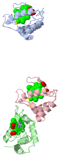
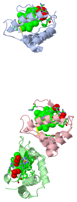
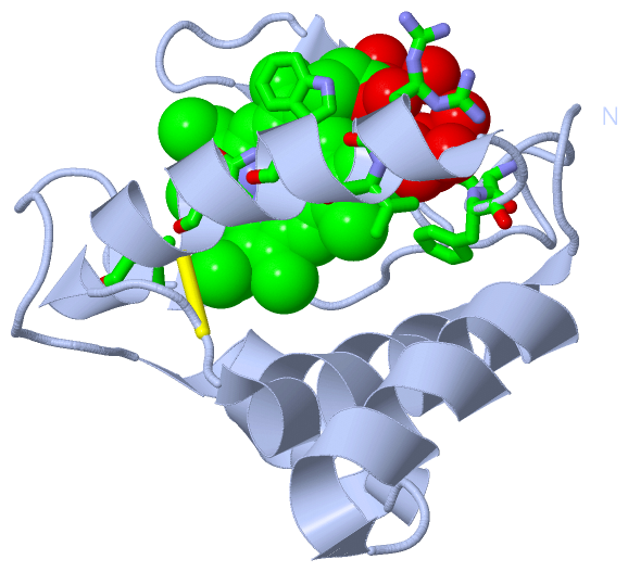
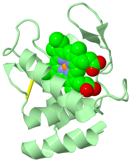
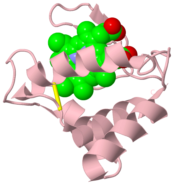
Asymmetric UnitChain A from PDB Type:PROTEIN Length:112 aligned with SHP_RHOS4 | P81238 from UniProtKB/Swiss-Prot Length:129 Alignment length:112 27 37 47 57 67 77 87 97 107 117 127 SHP_RHOS4 18 GDTSPAQLIAGYEAAAGAPADAERGRALFLSTQTGGKPDTPSCTTCHGADVTRAGQTRTGKEIAPLAPSATPDRFTDSARVEKWLGRNCNSVIGRDCTPGEKADLLAWLAAQ 129 SCOP domains d1dw3a_ A: SHP, an oxygen binding cytochrome c SCOP domains CATH domains 1dw3A00 A:1-112 Cytochrome c CATH domains Pfam domains ---------------------------------------------------------------------------------------------------------------- Pfam domains SAPs(SNPs) ---------------------------------------------------------------------------------------------------------------- SAPs(SNPs) PROSITE -------------------CYTC PDB: A:20-112 UniProt: 37-129 PROSITE Transcript ---------------------------------------------------------------------------------------------------------------- Transcript 1dw3 A 1 GDTSPAQLIAGYEAAAGAPADAERGRALFLSTQTGGKPDTPSCTTCHGADVTRAGQTRTGKEIAPLAPSATPDRFTDSARVEKWLGRNCNSVIGRDCTPGEKADLLAWLAAQ 112 10 20 30 40 50 60 70 80 90 100 110 Chain B from PDB Type:PROTEIN Length:112 aligned with SHP_RHOS4 | P81238 from UniProtKB/Swiss-Prot Length:129 Alignment length:112 27 37 47 57 67 77 87 97 107 117 127 SHP_RHOS4 18 GDTSPAQLIAGYEAAAGAPADAERGRALFLSTQTGGKPDTPSCTTCHGADVTRAGQTRTGKEIAPLAPSATPDRFTDSARVEKWLGRNCNSVIGRDCTPGEKADLLAWLAAQ 129 SCOP domains d1dw3b_ B: SHP, an oxygen binding cytochrome c SCOP domains CATH domains 1dw3B00 B:1-112 Cytochrome c CATH domains Pfam domains ---------------------------------------------------------------------------------------------------------------- Pfam domains SAPs(SNPs) ---------------------------------------------------------------------------------------------------------------- SAPs(SNPs) PROSITE -------------------CYTC PDB: B:20-112 UniProt: 37-129 PROSITE Transcript ---------------------------------------------------------------------------------------------------------------- Transcript 1dw3 B 1 GDTSPAQLIAGYEAAAGAPADAERGRALFLSTQTGGKPDTPSCTTCHGADVTRAGQTRTGKEIAPLAPSATPDRFTDSARVEKWLGRNCNSVIGRDCTPGEKADLLAWLAAQ 112 10 20 30 40 50 60 70 80 90 100 110 Chain C from PDB Type:PROTEIN Length:112 aligned with SHP_RHOS4 | P81238 from UniProtKB/Swiss-Prot Length:129 Alignment length:112 27 37 47 57 67 77 87 97 107 117 127 SHP_RHOS4 18 GDTSPAQLIAGYEAAAGAPADAERGRALFLSTQTGGKPDTPSCTTCHGADVTRAGQTRTGKEIAPLAPSATPDRFTDSARVEKWLGRNCNSVIGRDCTPGEKADLLAWLAAQ 129 SCOP domains d1dw3c_ C: SHP, an oxygen binding cytochrome c SCOP domains CATH domains 1dw3C00 C:1-112 Cytochrome c CATH domains Pfam domains ---------------------------------------------------------------------------------------------------------------- Pfam domains SAPs(SNPs) ---------------------------------------------------------------------------------------------------------------- SAPs(SNPs) PROSITE -------------------CYTC PDB: C:20-112 UniProt: 37-129 PROSITE Transcript ---------------------------------------------------------------------------------------------------------------- Transcript 1dw3 C 1 GDTSPAQLIAGYEAAAGAPADAERGRALFLSTQTGGKPDTPSCTTCHGADVTRAGQTRTGKEIAPLAPSATPDRFTDSARVEKWLGRNCNSVIGRDCTPGEKADLLAWLAAQ 112 10 20 30 40 50 60 70 80 90 100 110
|
||||||||||||||||||||
SCOP Domains (1, 3)
Asymmetric Unit
 |
CATH Domains (1, 3)
Asymmetric Unit
|
Pfam Domains (0, 0)| (no "Pfam Domain" information available for 1DW3) |
Gene Ontology (4, 4)|
Asymmetric Unit(hide GO term definitions) Chain A,B,C (SHP_RHOS4 | P81238)
|
||||||||||||||||||||||||||||||||||||
Interactive Views
|
||||||||||||||||||||||||||||||||||||||||||||||||||||||||||||||||||||||||||||||||||||||||||||||||||||||||||||||||||||||||||||||||||||||||||||||||||||||||||||||||
Still Images
|
||||||||||||||||
Databases
|
||||||||||||||||||||||||||||||||||||||||||||||||||||||||||||||||||||||||||||||||||||||||||||||||||||||||||||||||||||||||||||||||||||||||||||||||||||||||||||||||
Analysis Tools
|
|||||||||||||||||||||||||||||||||||||||||||||||||||||||||||||
Entries Sharing at Least One Protein Chain (UniProt ID)
Related Entries Specified in the PDB File
|
|