| molecular function |
|---|
| | GO:0003677 | | DNA binding | | Any molecular function by which a gene product interacts selectively and non-covalently with DNA (deoxyribonucleic acid). |
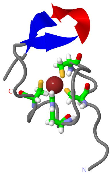
| | GO:0046872 | | metal ion binding | | Interacting selectively and non-covalently with any metal ion. |
| | GO:0003676 | | nucleic acid binding | | Interacting selectively and non-covalently with any nucleic acid. |
| | GO:0000166 | | nucleotide binding | | Interacting selectively and non-covalently with a nucleotide, any compound consisting of a nucleoside that is esterified with (ortho)phosphate or an oligophosphate at any hydroxyl group on the ribose or deoxyribose. |
| | GO:0039660 | | structural constituent of virion | | The action of a molecule that contributes to the structural integrity of a virion. |
| | GO:0005198 | | structural molecule activity | | The action of a molecule that contributes to the structural integrity of a complex or its assembly within or outside a cell. |
| | GO:0008270 | | zinc ion binding | | Interacting selectively and non-covalently with zinc (Zn) ions. |
| biological process |
|---|
| | GO:0046755 | | viral budding | | A viral process by which enveloped viruses acquire a host-derived membrane enriched in viral proteins to form their external envelope. The process starts when nucleocapsids, assembled or in the process of being built, induce formation of a membrane curvature in the host plasma or organelle membrane and wrap up in the forming bud. The process ends when the bud is eventually pinched off by membrane scission to release the enveloped particle into the lumenal or extracellular space. |
| | GO:0039702 | | viral budding via host ESCRT complex | | Viral budding which uses a host ESCRT protein complex, or complexes, to mediate the budding process. |
| | GO:0016032 | | viral process | | A multi-organism process in which a virus is a participant. The other participant is the host. Includes infection of a host cell, replication of the viral genome, and assembly of progeny virus particles. In some cases the viral genetic material may integrate into the host genome and only subsequently, under particular circumstances, 'complete' its life cycle. |
| | GO:0019076 | | viral release from host cell | | The dissemination of mature viral particles from the host cell, e.g. by cell lysis or the budding of virus particles from the cell membrane. |
| cellular component |
|---|
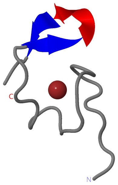
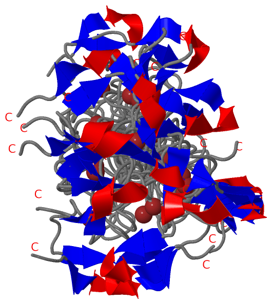
| | GO:0019028 | | viral capsid | | The protein coat that surrounds the infective nucleic acid in some virus particles. It comprises numerous regularly arranged subunits, or capsomeres. |
| | GO:0019013 | | viral nucleocapsid | | The complete protein-nucleic acid complex that is the packaged form of the genome in a virus particle. |
| | GO:0019012 | | virion | | The complete fully infectious extracellular virus particle. |
 Description