Show PDB file:   
         Plain Text   HTML   (compressed file size)
QuickSearch:   
by PDB,NDB,UniProt,PROSITE Code or Search Term(s)  
(-)Biol.Unit 1 - manually
(-)Asymmetric Unit
(-)Asym. Unit - sites
(-)Biological Unit 1
(-)Biol. Unit 1 - sites
collapse expand < >
Image Biol.Unit 1 - manually
Biol.Unit 1 - manually  (Jmol Viewer)
Image Asymmetric Unit
Asymmetric Unit  (Jmol Viewer)
Image Asym. Unit - sites
Asym. Unit - sites  (Jmol Viewer)
Image Biological Unit 1
Biological Unit 1  (Jmol Viewer)
Image Biol. Unit 1 - sites
Biol. Unit 1 - sites  (Jmol Viewer)

(-) Description

Title :  2FE-2S FERREDOXIN FROM HALOARCULA MARISMORTUI
 
Authors :  F. Frolow, M. Harel, J. L. Sussman, M. Shoham
Date :  08 Apr 96  (Deposition) - 01 Aug 96  (Release) - 24 Feb 09  (Revision)
Method :  X-RAY DIFFRACTION
Resolution :  1.90
Chains :  Asym. Unit :  A
Biol. Unit 1:  A  (6x)
Keywords :  Halophilic Protein, Redox Protein, Iron-Sulfur, Electron Transport (Keyword Search: [Gene Ontology, PubMed, Web (Google))
 
Reference :  F. Frolow, M. Harel, J. L. Sussman, M. Mevarech, M. Shoham
Insights Into Protein Adaptation To A Saturated Salt Environment From The Crystal Structure Of A Halophilic 2Fe-2S Ferredoxin.
Nat. Struct. Biol. V. 3 452 1996
PubMed-ID: 8612076  |  Reference-DOI: 10.1038/NSB0596-452
(for further references see the PDB file header)

(-) Compounds

Molecule 1 - 2FE-2S FERREDOXIN
    ChainsA
    Organism ScientificHALOARCULA MARISMORTUI
    Organism Taxid2238

 Structural Features

(-) Chains, Units

  1
Asymmetric Unit A
Biological Unit 1 (6x)A

Summary Information (see also Sequences/Alignments below)
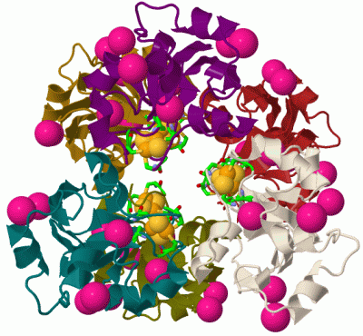
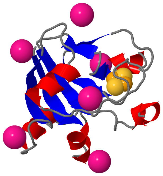
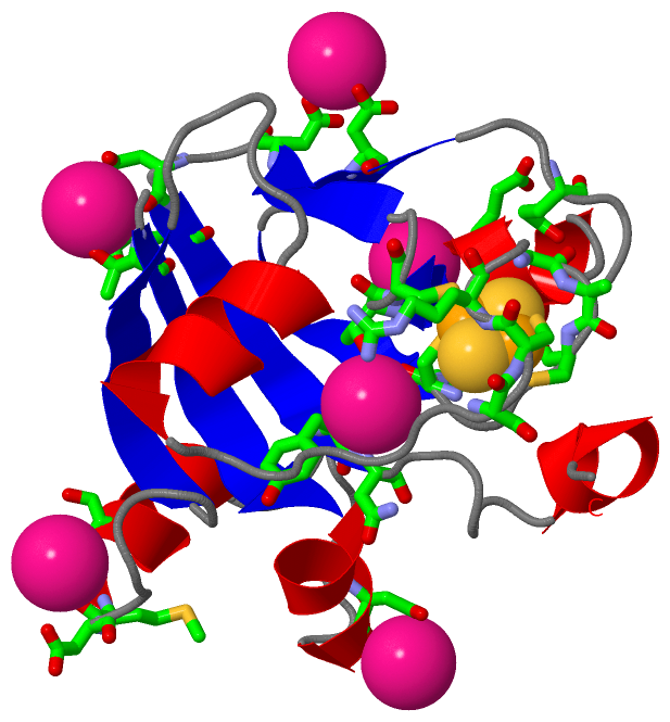
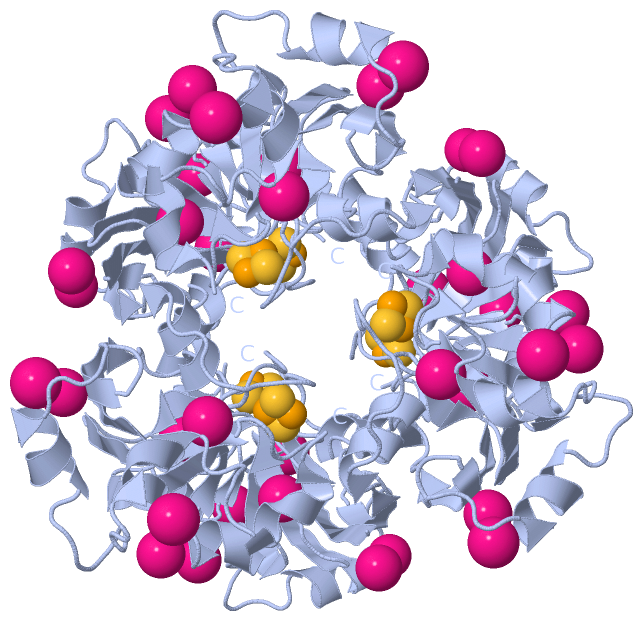
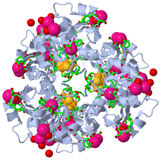
(-) Ligands, Modified Residues, Ions  (2, 7)

Asymmetric Unit (2, 7)
No.NameCountTypeFull Name
1FES1Ligand/IonFE2/S2 (INORGANIC) CLUSTER
2K6Ligand/IonPOTASSIUM ION
Biological Unit 1 (1, 6)
No.NameCountTypeFull Name
1FES6Ligand/IonFE2/S2 (INORGANIC) CLUSTER
2K-1Ligand/IonPOTASSIUM ION

(-) Sites  (8, 8)

Asymmetric Unit (8, 8)
No.NameEvidenceResiduesDescription
122UNKNOWNCYS A:63 , CYS A:68 , CYS A:71 , CYS A:102FE2S2 IRON-SULFUR REDOX CENTER.
2AC1SOFTWAREASP A:12 , ASP A:31 , HOH A:418BINDING SITE FOR RESIDUE K A 201
3AC2SOFTWAREASP A:81 , GLN A:85 , ASP A:107 , HOH A:274 , HOH A:426BINDING SITE FOR RESIDUE K A 202
4AC3SOFTWARESER A:28 , MET A:30 , HOH A:374BINDING SITE FOR RESIDUE K A 203
5AC4SOFTWARECYS A:71 , ALA A:72 , THR A:101 , TYR A:115 , ASN A:116 , HOH A:211BINDING SITE FOR RESIDUE K A 204
6AC5SOFTWAREGLU A:94 , ASN A:97 , HOH A:214 , HOH A:439 , HOH A:440BINDING SITE FOR RESIDUE K A 205
7AC6SOFTWARETHR A:2 , ASP A:109 , GLU A:110BINDING SITE FOR RESIDUE K A 206
8AC7SOFTWARESER A:62 , CYS A:63 , ARG A:64 , GLY A:66 , ALA A:67 , CYS A:68 , CYS A:71 , CYS A:102BINDING SITE FOR RESIDUE FES A 129

(-) SS Bonds  (0, 0)

(no "SS Bond" information available for 1DOI)

(-) Cis Peptide Bonds  (0, 0)

(no "Cis Peptide Bond" information available for 1DOI)

 Sequence-Structure Mapping

(-) SAPs(SNPs)/Variants  (0, 0)

(no "SAP(SNP)/Variant" information available for 1DOI)

(-) PROSITE Motifs  (2, 2)

Asymmetric Unit (2, 2)
 PROSITEUniProtKBPDB
No.IDACDescriptionIDLocationCountLocation
12FE2S_FER_2PS51085 2Fe-2S ferredoxin-type iron-sulfur binding domain profile.FER1_HALMA29-120  1A:28-119
22FE2S_FER_1PS00197 2Fe-2S ferredoxin-type iron-sulfur binding region signature.FER1_HALMA64-72  1A:63-71
Biological Unit 1 (2, 12)
 PROSITEUniProtKBPDB
No.IDACDescriptionIDLocationCountLocation
12FE2S_FER_2PS51085 2Fe-2S ferredoxin-type iron-sulfur binding domain profile.FER1_HALMA29-120  6A:28-119
22FE2S_FER_1PS00197 2Fe-2S ferredoxin-type iron-sulfur binding region signature.FER1_HALMA64-72  6A:63-71

(-) Exons   (0, 0)

(no "Exon" information available for 1DOI)

(-) Sequences/Alignments

Asymmetric Unit
   Reformat: Number of residues per line =  ('0' or empty: single-line sequence representation)
  Number of residues per labelling interval =   
  UniProt sequence: complete  aligned part    
   Show mapping: SCOP domains CATH domains Pfam domains Secondary structure (by author)
SAPs(SNPs) PROSITE motifs Exons
(details for a mapped element are shown in a popup box when the mouse pointer rests over it)
Chain A from PDB  Type:PROTEIN  Length:128
 aligned with FER1_HALMA | P00217 from UniProtKB/Swiss-Prot  Length:129

    Alignment length:128
                                    11        21        31        41        51        61        71        81        91       101       111       121        
           FER1_HALMA     2 PTVEYLNYEVVDDNGWDMYDDDVFGEASDMDLDDEDYGSLEVNEGEYILEAAEAQGYDWPFSCRAGACANCAAIVLEGDIDMDMQQILSDEEVEDKNVRLTCIGSPDADEVKIVYNAKHLDYLQNRVI 129
               SCOP domains d1doia_ A: 2Fe-2S ferredoxin                                                                                                     SCOP domains
               CATH domains 1doiA00 A:1-128  [code=3.10.20.30, no name defined]                                                                              CATH domains
               Pfam domains -------------------------------------------------------------------------------------------------------------------------------- Pfam domains
         Sec.struct. author .eeeeeehhhhhh.........hhhhhhh.......eeeee......hhhhhhhh................eeeeee..eee.......hhhhh...eee...eee...eeeeee.....hhhhhh.. Sec.struct. author
                 SAPs(SNPs) -------------------------------------------------------------------------------------------------------------------------------- SAPs(SNPs)
                PROSITE (1) ---------------------------2FE2S_FER_2  PDB: A:28-119 UniProt: 29-120                                                  --------- PROSITE (1)
                PROSITE (2) --------------------------------------------------------------2FE2S_FER--------------------------------------------------------- PROSITE (2)
                 Transcript -------------------------------------------------------------------------------------------------------------------------------- Transcript
                 1doi A   1 PTVEYLNYEVVDDNGWDMYDDDVFGEASDMDLDDEDYGSLEVNEGEYILEAAEAQGYDWPFSCRAGACANCAAIVLEGDIDMDMQQILSDEEVEDKNVRLTCIGSPDADEVKIVYNAKHLDYLQNRVI 128
                                    10        20        30        40        50        60        70        80        90       100       110       120        

   Legend:   → Mismatch (orange background)
  - → Gap (green background, '-', border residues have a numbering label)
    → Modified Residue (blue background, lower-case, 'x' indicates undefined single-letter code, labelled with number + name)
  x → Chemical Group (purple background, 'x', labelled with number + name, e.g. ACE or NH2)
  extra numbering lines below/above indicate numbering irregularities and modified residue names etc., number ends below/above '|'

 Classification and Annotation

(-) SCOP Domains  (1, 1)

Asymmetric Unit

(-) CATH Domains  (1, 1)

Asymmetric Unit
(-)
Class: Alpha Beta (26913)

(-) Pfam Domains  (0, 0)

(no "Pfam Domain" information available for 1DOI)

(-) Gene Ontology  (5, 5)

Asymmetric Unit(hide GO term definitions)
Chain A   (FER1_HALMA | P00217)
molecular function
    GO:0051537    2 iron, 2 sulfur cluster binding    Interacting selectively and non-covalently with a 2 iron, 2 sulfur (2Fe-2S) cluster; this cluster consists of two iron atoms, with two inorganic sulfur atoms found between the irons and acting as bridging ligands.
    GO:0009055    electron carrier activity    Any molecular entity that serves as an electron acceptor and electron donor in an electron transport chain. An electron transport chain is a process in which a series of electron carriers operate together to transfer electrons from donors to any of several different terminal electron acceptors to generate a transmembrane electrochemical gradient.
    GO:0051536    iron-sulfur cluster binding    Interacting selectively and non-covalently with an iron-sulfur cluster, a combination of iron and sulfur atoms.
    GO:0046872    metal ion binding    Interacting selectively and non-covalently with any metal ion.
biological process
    GO:0055114    oxidation-reduction process    A metabolic process that results in the removal or addition of one or more electrons to or from a substance, with or without the concomitant removal or addition of a proton or protons.

 Visualization

(-) Interactive Views

Asymmetric Unit
  Complete Structure
    Jena3D(integrated viewing of ligand, site, SAP, PROSITE, SCOP information)
    WebMol | AstexViewer[tm]@PDBe
(Java Applets, require no local installation except for Java; loading may be slow)
    STRAP
(Java WebStart application, automatic local installation, requires Java; full application with system access!)
    RasMol
(require local installation)
    Molscript (VRML)
(requires installation of a VRML viewer; select preferred view via VRML and generate a mono or stereo PDF format file)
 
  Ligands, Modified Residues, Ions
    FES  [ RasMol | Jena3D ]  +environment [ RasMol | Jena3D ]
    K  [ RasMol | Jena3D ]  +environment [ RasMol | Jena3D ]
 
  Sites
    22  [ RasMol ]  +environment [ RasMol ]
    AC1  [ RasMol ]  +environment [ RasMol ]
    AC2  [ RasMol ]  +environment [ RasMol ]
    AC3  [ RasMol ]  +environment [ RasMol ]
    AC4  [ RasMol ]  +environment [ RasMol ]
    AC5  [ RasMol ]  +environment [ RasMol ]
    AC6  [ RasMol ]  +environment [ RasMol ]
    AC7  [ RasMol ]  +environment [ RasMol ]
 
  Cis Peptide Bonds
(no "Cis Peptide Bonds" information available for 1doi)
 
Biological Unit
  Complete Structure
    Biological Unit 1  [ Jena3D ]

(-) Still Images

Jmol
  protein: cartoon or spacefill or dots and stick; nucleic acid: cartoon and stick; ligands: spacefill; active site: stick
Molscript
  protein, nucleic acid: cartoon; ligands: spacefill; active site: ball and stick

 Databases and Analysis Tools

(-) Databases

Access by PDB/NDB ID
  1doi
    Family and Domain InformationProDom | SYSTERS
    General Structural InformationGlycoscienceDB | MMDB | NDB | OCA | PDB | PDBe | PDBj | PDBsum | PDBWiki | PQS | PROTEOPEDIA
    Orientation in MembranesOPM
    Protein SurfaceSURFACE
    Secondary StructureDSSP (structure derived) | HSSP (homology derived)
    Structural GenomicsGeneCensus
    Structural NeighboursCE | VAST
    Structure ClassificationCATH | Dali | SCOP
    Validation and Original DataBMRB Data View | BMRB Restraints Grid | EDS | PROCHECK | RECOORD | WHAT_CHECK
 
Access by UniProt ID/Accession number
  FER1_HALMA | P00217
    Comparative Protein Structure ModelsModBase
    Genomic InformationEnsembl
    Protein-protein InteractionDIP
    Sequence, Family and Domain InformationInterPro | Pfam | SMART | UniProtKB/SwissProt
 
Access by Enzyme Classificator   (EC Number)
  (no 'Enzyme Classificator' available)
    General Enzyme InformationBRENDA | EC-PDB | Enzyme | IntEnz
    PathwayKEGG | MetaCyc
 
Access by Disease Identifier   (MIM ID)
  (no 'MIM ID' available)
    Disease InformationOMIM
 
Access by GenAge ID
  (no 'GenAge ID' available)
    Age Related InformationGenAge

(-) Analysis Tools

Access by PDB/NDB ID
    Domain InformationXDom
    Interatomic Contacts of Structural UnitsCSU
    Ligand-protein ContactsLPC
    Protein CavitiescastP
    Sequence and Secondary StructurePDBCartoon
    Structure AlignmentSTRAP(Java WebStart application, automatic local installation, requires Java; full application with system access!)
    Structure and Sequence BrowserSTING
 
Access by UniProt ID/Accession number
  FER1_HALMA | P00217
    Protein Disorder PredictionDisEMBL | FoldIndex | GLOBPLOT (for more information see DisProt)

 Related Entries

(-) Entries Sharing at Least One Protein Chain (UniProt ID)

(no "Entries Sharing at Least One Protein Chain" available for 1DOI)

(-) Related Entries Specified in the PDB File

(no "Related Entries Specified in the PDB File" available for 1DOI)