|
|
|
|
 Description
|
|
Compounds
|
||||||||||||||||||||||||||||||||||||
Chains, Units
Summary Information (see also Sequences/Alignments below) |
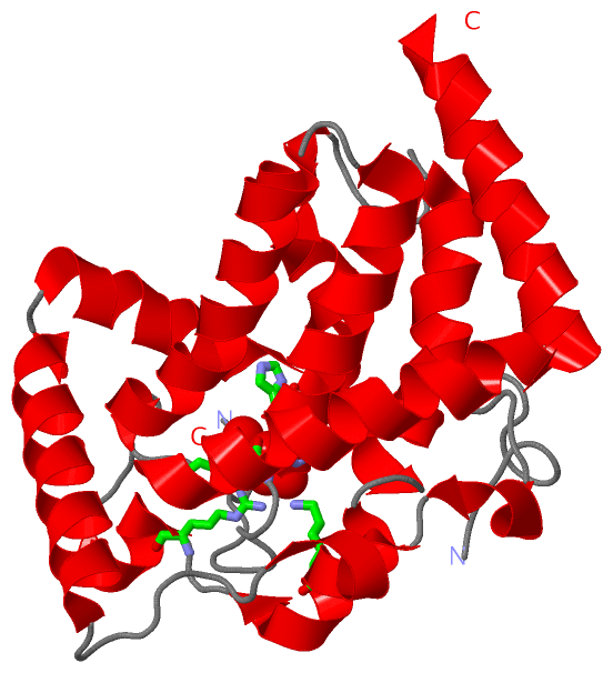
Ligands, Modified Residues, Ions (1, 1)
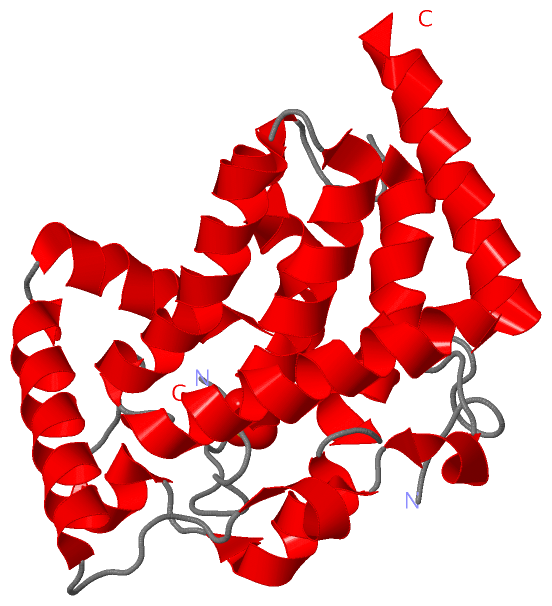
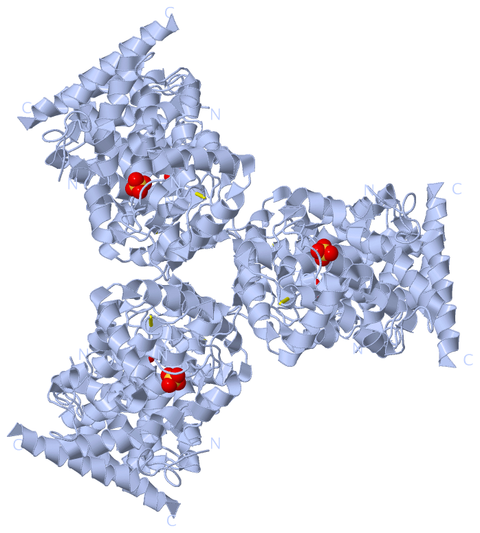
Asymmetric Unit (1, 1)
|
Sites (1, 1)
Asymmetric Unit (1, 1)
|
SS Bonds (1, 1)
Asymmetric Unit
|
||||||||
Cis Peptide Bonds (1, 1)
Asymmetric Unit
|
||||||||
SAPs(SNPs)/Variants (0, 0)| (no "SAP(SNP)/Variant" information available for 1D2T) |
PROSITE Motifs (0, 0)| (no "PROSITE Motif" information available for 1D2T) |
Exons (0, 0)| (no "Exon" information available for 1D2T) |
Sequences/Alignments
Asymmetric UnitChain A from PDB Type:PROTEIN Length:222 aligned with Q9S1A6_SHIBL | Q9S1A6 from UniProtKB/TrEMBL Length:249 Alignment length:224 34 44 54 64 74 84 94 104 114 124 134 144 154 164 174 184 194 204 214 224 234 244 Q9S1A6_SHIBL 25 GNDTTTKPDLYYLKNSEAINSLALLPPPPAVGSIAFLNDQAMYEQGRLLRNTERGKLAAEDANLSSGGVANAFSGAFGSPITEKDAPALHKLLTNMIEDAGDLATRSAKDHYMRIRPFAFYGVSTCNTTEQDKLSKNGSYPSGHTSIGWATALVLAEINPQRQNEILKRGYELGQSRVICGYHWQSDVDAARVVGSAVVATLHTNPAFQQQLQKAKAEFAQHQK 248 SCOP domains d1d2ta_ A: Bacterial acid phosphatase SCOP domains CATH domains 1d2tA00 A:7-230 Vanadium-containing Chloroperoxidase, domain 1 CATH domains Pfam domains -------------------------------------------------------------------------------------------------------------------------------------------------------------------------------------------------------------------------------- Pfam domains SAPs(SNPs) -------------------------------------------------------------------------------------------------------------------------------------------------------------------------------------------------------------------------------- SAPs(SNPs) PROSITE -------------------------------------------------------------------------------------------------------------------------------------------------------------------------------------------------------------------------------- PROSITE Transcript -------------------------------------------------------------------------------------------------------------------------------------------------------------------------------------------------------------------------------- Transcript 1d2t A 7 GNDTTTKPDLYYLKNSEAINSLALLPPPPAVGSIAFLNDQAMYEQGRLLRNTERGKLAAEDANLSSGGVANAFSGAFGSPITEKDAPALHKLLTNMIEDAGDLATRSAKDHYMRIRPFAFYGVSTCNT--QDKLSKNGSYPSGHTSIGWATALVLAEINPQRQNEILKRGYELGQSRVICGYHWQSDVDAARVVGSAVVATLHTNPAFQQQLQKAKAEFAQHQK 230 16 26 36 46 56 66 76 86 96 106 116 126 | -| 146 156 166 176 186 196 206 216 226 134 | 137
|
||||||||||||||||||||
SCOP Domains (1, 1)
Asymmetric Unit
 |
CATH Domains (1, 1)
Asymmetric Unit
|
Pfam Domains (0, 0)| (no "Pfam Domain" information available for 1D2T) |
Gene Ontology (4, 4)|
Asymmetric Unit(hide GO term definitions) Chain A (Q9S1A6_SHIBL | Q9S1A6)
|
||||||||||||||||||||||||||||||||||||||||||
Interactive Views
|
|||||||||||||||||||||||||||||||||||||||||||||||||||||||||||||||||||||||||||||||||||||||||||||||||||||||||||||||||||||||||||||||||||||||||
Still Images
|
||||||||||||||||
Databases
|
||||||||||||||||||||||||||||||||||||||||||||||||||||||||||||||||||||||||||||||||||||||||||||||||||||||||||||||||||||||||||||||||||||||||||||||||||||||||||||||||
Analysis Tools
|
|||||||||||||||||||||||||||||||||||||||||||||||||||||||||||||
Entries Sharing at Least One Protein Chain (UniProt ID)
Related Entries Specified in the PDB File
|
|