|
|
|
|
 Description
|
|
Compounds
|
||||||||||||||||
Chains, Units
Summary Information (see also Sequences/Alignments below) |
Ligands, Modified Residues, Ions (3, 12)
Asymmetric Unit (3, 12)
|
Sites (0, 0)| (no "Site" information available for 1CS7) |
SS Bonds (0, 0)| (no "SS Bond" information available for 1CS7) |
Cis Peptide Bonds (0, 0)| (no "Cis Peptide Bond" information available for 1CS7) |
SAPs(SNPs)/Variants (0, 0)| (no "SAP(SNP)/Variant" information available for 1CS7) |
PROSITE Motifs (0, 0)| (no "PROSITE Motif" information available for 1CS7) |
Exons (0, 0)| (no "Exon" information available for 1CS7) |
Sequences/Alignments
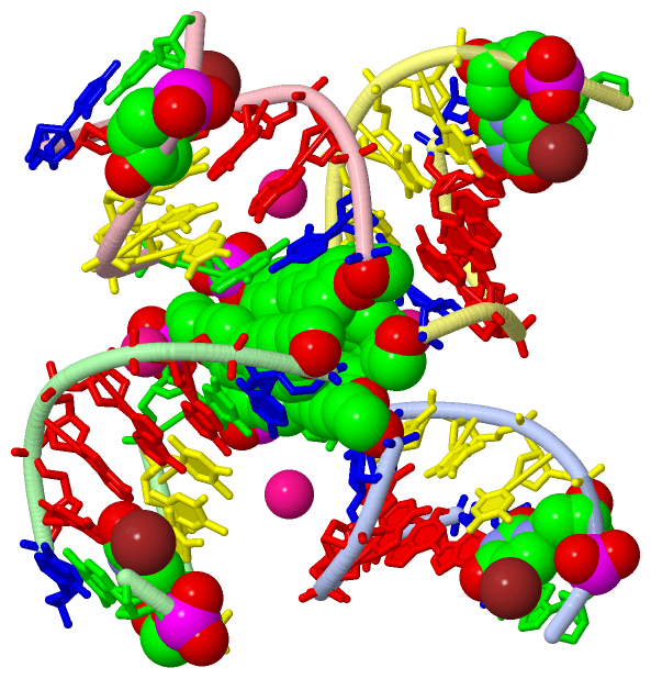
Asymmetric Unit
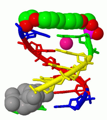
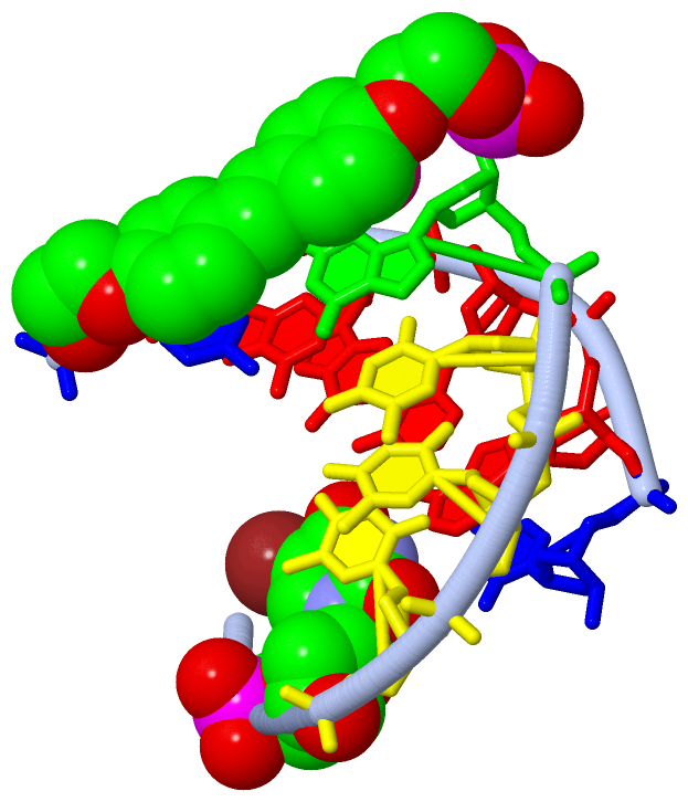
Chain A from PDB  Type:DNA  Length:13

                 1cs7 A   1 GuTTTGxCAAAAC  14
                             |    ||11
                             |    ||
                             2-BRU||
                                  7-S02
                                   9
Chain B from PDB  Type:DNA  Length:13

                 1cs7 B 101 GuTTTGxCAAAAC 114
                             |    |111
                             |    ||
                           102-BRU||
                                107-S02
                                 109
Chain C from PDB  Type:DNA  Length:13

                 1cs7 C 201 GuTTTGxCAAAAC 214
                             |    |211
                             |    ||
                           202-BRU||
                                207-S02
                                 209
Chain D from PDB  Type:DNA  Length:13

                 1cs7 D 301 GuTTTGxCAAAAC 314
                             |    |311
                             |    ||
                           302-BRU||
                                307-S02
                                 309
|
||||||||||||||||||||
SCOP Domains (0, 0)| (no "SCOP Domain" information available for 1CS7) |
CATH Domains (0, 0)| (no "CATH Domain" information available for 1CS7) |
Pfam Domains (0, 0)| (no "Pfam Domain" information available for 1CS7) |
Gene Ontology (0, 0)|
Asymmetric Unit(hide GO term definitions)
(no "Gene Ontology" information available for 1CS7)
|
Interactive Views
|
||||||||||||||||||||||||||||||||||||||||||||||||||||||||||||||||||||||||||||||||||||||||||||||||||||||||||||||||||||||||||||||||||||||||||||||||||||||||||||||||||||
Still Images
|
||||||||||||||||
Databases
|
||||||||||||||||||||||||||||||||||||||||||||||||||||||||||||||||||||||||||||||||||||||||||||||||||||||||||||||||||||||||||||||||||||||||||||||||||||||||||||||||
Analysis Tools
|
|||||||||||||||||||||||||||||||||||||||||||||||||||||||||||||
Entries Sharing at Least One Protein Chain (UniProt ID)
Related Entries Specified in the PDB File
|
|