|
|
|
|
 Description
|
|
Compounds
|
||||||||||||||||||||||||
Chains, Units
Summary Information (see also Sequences/Alignments below) |
Ligands, Modified Residues, Ions (1, 1)
Asymmetric Unit (1, 1)
|
Sites (1, 1)
Asymmetric Unit (1, 1)
|
SS Bonds (0, 0)| (no "SS Bond" information available for 1BVQ) |
Cis Peptide Bonds (0, 0)| (no "Cis Peptide Bond" information available for 1BVQ) |
SAPs(SNPs)/Variants (0, 0)| (no "SAP(SNP)/Variant" information available for 1BVQ) |
PROSITE Motifs (1, 1)
Asymmetric Unit (1, 1)
|
||||||||||||||||||||||||||||||||||||||||||||||||
Exons (0, 0)| (no "Exon" information available for 1BVQ) |
Sequences/Alignments
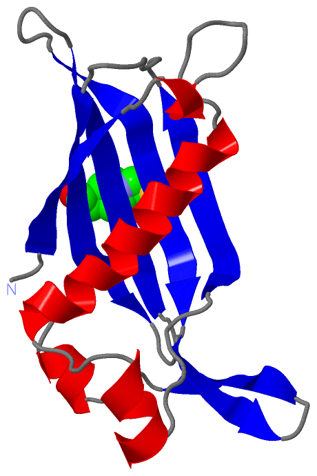
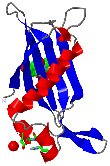
Asymmetric UnitChain A from PDB Type:PROTEIN Length:139 aligned with 4HBT_PSEUC | P56653 from UniProtKB/Swiss-Prot Length:141 Alignment length:139 11 21 31 41 51 61 71 81 91 101 111 121 131 4HBT_PSEUC 2 ARSITMQQRIEFGDCDPAGIVWFPNYHRWLDAASRNYFIKCGLPPWRQTVVERGIVGTPIVSCNASFVCTASYDDVLTIETCIKEWRRKSFVQRHSVSRTTPGGDVQLVMRADEIRVFAMNDGERLRAIEVPADYIELC 140 SCOP domains d1bvqa_ A: 4-hydroxybenzoyl-CoA thioesterase SCOP domains CATH domains 1bvqA00 A:2-140 [code=3.10.129.10, no name defined] CATH domains Pfam domains ------------------------------------------------------------------------------------------------------------------------------------------- Pfam domains SAPs(SNPs) ------------------------------------------------------------------------------------------------------------------------------------------- SAPs(SNPs) PROSITE --------4HBCOA_THIOESTERAS----------------------------------------------------------------------------------------------------------------- PROSITE Transcript ------------------------------------------------------------------------------------------------------------------------------------------- Transcript 1bvq A 2 ARSITMQQRIEFGDCDPAGIVWYPNYHRWLDAASRNYFIKCGLPPWRQTVVERGIVGTPIVSCNASFVCTASYDDVLTIETCIKEWRRKSFVQRHSVSRTTPGGDVQLVMRADEIRVFAMNDGERLRAIEVPADYIELC 140 11 21 31 41 51 61 71 81 91 101 111 121 131
|
||||||||||||||||||||
SCOP Domains (1, 1)
Asymmetric Unit
 |
CATH Domains (1, 1)
Asymmetric Unit
|
Pfam Domains (0, 0)| (no "Pfam Domain" information available for 1BVQ) |
Gene Ontology (2, 2)|
Asymmetric Unit(hide GO term definitions) Chain A (4HBT_PSEUC | P56653)
|
||||||||||||||||||
Interactive Views
|
||||||||||||||||||||||||||||||||||||||||||||||||||||||||||||||||||||||||||||||||||||||||||||||||||||||||||||||||||||||||||||||||||||||||
Still Images
|
||||||||||||||||
Databases
|
||||||||||||||||||||||||||||||||||||||||||||||||||||||||||||||||||||||||||||||||||||||||||||||||||||||||||||||||||||||||||||||||||||||||||||||||||||||||||||||||
Analysis Tools
|
|||||||||||||||||||||||||||||||||||||||||||||||||||||||||||||
Entries Sharing at Least One Protein Chain (UniProt ID)
Related Entries Specified in the PDB File
|
|