Show PDB file:   
         Plain Text   HTML   (compressed file size)
QuickSearch:   
by PDB,NDB,UniProt,PROSITE Code or Search Term(s)  
(-)Asym./Biol. Unit - manually
(-)Asym./Biol. Unit
(-)Asym./Biol. Unit - sites
collapse expand < >
Image Asym./Biol. Unit - manually
Asym./Biol. Unit - manually  (Jmol Viewer)
Image Asym./Biol. Unit
Asym./Biol. Unit  (Jmol Viewer)
Image Asym./Biol. Unit - sites
Asym./Biol. Unit - sites  (Jmol Viewer)

(-) Description

Title :  COLLAGEN
 
Authors :  R. Z. Kramer, J. Bella, P. Mayville, B. Brodsky, H. M. Berman
Date :  13 Jul 98  (Deposition) - 16 Feb 99  (Release) - 22 Dec 10  (Revision)
Method :  X-RAY DIFFRACTION
Resolution :  2.00
Chains :  Asym./Biol. Unit :  A,B,C
Keywords :  Collagen, Hydroxyproline, Hydrogen Bonding, Triple Helix, Type Iii Collagen, Structural Protein (Keyword Search: [Gene Ontology, PubMed, Web (Google))
 
Reference :  R. Z. Kramer, J. Bella, P. Mayville, B. Brodsky, H. M. Berman
Sequence Dependent Conformational Variations Of Collagen Triple-Helical Structure.
Nat. Struct. Biol. V. 6 454 1999
PubMed-ID: 10331873  |  Reference-DOI: 10.1038/8259

(-) Compounds

Molecule 1 - T3-785
    ChainsA, B, C
    EngineeredYES
    Other DetailsSYNTHETIC PEPTIDE CONTAINS REGION FROM HUMAN TYPE III COLLAGEN

 Structural Features

(-) Chains, Units

  123
Asymmetric/Biological Unit ABC

Summary Information (see also Sequences/Alignments below)

(-) Ligands, Modified Residues, Ions  (2, 26)

Asymmetric/Biological Unit (2, 26)
No.NameCountTypeFull Name
1ACY5Ligand/IonACETIC ACID
2HYP21Mod. Amino Acid4-HYDROXYPROLINE

(-) Sites  (5, 5)

Asymmetric Unit (5, 5)
No.NameEvidenceResiduesDescription
1AC1SOFTWAREARG B:44 , HOH B:112 , HOH B:125 , ACY B:405BINDING SITE FOR RESIDUE ACY B 401
2AC2SOFTWAREARG A:14 , ILE C:70 , THR C:71 , HOH C:145 , HOH C:195BINDING SITE FOR RESIDUE ACY C 402
3AC3SOFTWAREHOH B:111 , ACY B:404 , HOH C:117 , HOH C:210BINDING SITE FOR RESIDUE ACY B 403
4AC4SOFTWARELEU A:16 , GLY B:51 , PRO B:52 , HYP B:53 , HOH B:119 , ACY B:403BINDING SITE FOR RESIDUE ACY B 404
5AC5SOFTWAREALA B:43 , ARG B:44 , HOH B:125 , HOH B:131 , ACY B:401BINDING SITE FOR RESIDUE ACY B 405

(-) SS Bonds  (0, 0)

(no "SS Bond" information available for 1BKV)

(-) Cis Peptide Bonds  (1, 1)

Asymmetric/Biological Unit
No.Residues
1Pro B:58 -Hyp B:59

 Sequence-Structure Mapping

(-) SAPs(SNPs)/Variants  (0, 0)

(no "SAP(SNP)/Variant" information available for 1BKV)

(-) PROSITE Motifs  (0, 0)

(no "PROSITE Motif" information available for 1BKV)

(-) Exons   (0, 0)

(no "Exon" information available for 1BKV)

(-) Sequences/Alignments

Asymmetric/Biological Unit
   Reformat: Number of residues per line =  ('0' or empty: single-line sequence representation)
  Number of residues per labelling interval =   
  UniProt sequence: complete  aligned part    
   Show mapping: SCOP domains CATH domains Pfam domains Secondary structure (by author)
SAPs(SNPs) PROSITE motifs Exons
(details for a mapped element are shown in a popup box when the mouse pointer rests over it)
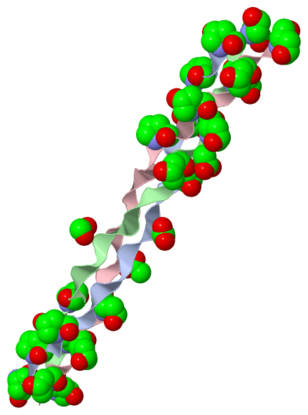
Chain A from PDB  Type:PROTEIN  Length:29
                                                            
               SCOP domains d1bkva_ A:                    SCOP domains
               CATH domains ----------------------------- CATH domains
               Pfam domains ----------------------------- Pfam domains
         Sec.struct. author ............................. Sec.struct. author
                 SAPs(SNPs) ----------------------------- SAPs(SNPs)
                    PROSITE ----------------------------- PROSITE
                 Transcript ----------------------------- Transcript
                  1bkv A  2 pGPpGPpGITGARGLAGPpGPpGPpGPpG 30
                            |  |  | 11        21 |  |  | 
                            |  |  |          20-HYP |  | 
                            2-HYP |             23-HYP | 
                               5-HYP               26-HYP
                                  8-HYP               29-HYP

Chain B from PDB  Type:PROTEIN  Length:30
                                                             
               SCOP domains d1bkvb_ B:                     SCOP domains
               CATH domains ------------------------------ CATH domains
               Pfam domains ------------------------------ Pfam domains
         Sec.struct. author .............................. Sec.struct. author
                 SAPs(SNPs) ------------------------------ SAPs(SNPs)
                    PROSITE ------------------------------ PROSITE
                 Transcript ------------------------------ Transcript
                  1bkv B 31 PpGPpGPpGITGARGLAGPpGPpGPpGPpG 60
                             |  |  |40        50  |  |  60
                             |  |  |          50-HYP |  | 
                            32-HYP |             53-HYP | 
                               35-HYP               56-HYP
                                  38-HYP               59-HYP

Chain C from PDB  Type:PROTEIN  Length:30
                                                             
               SCOP domains d1bkvc_ C:                     SCOP domains
               CATH domains ------------------------------ CATH domains
               Pfam domains ------------------------------ Pfam domains
         Sec.struct. author .............................. Sec.struct. author
                 SAPs(SNPs) ------------------------------ SAPs(SNPs)
                    PROSITE ------------------------------ PROSITE
                 Transcript ------------------------------ Transcript
                  1bkv C 61 PpGPpGPpGITGARGLAGPpGPpGPpGPpG 90
                             |  |  |70        80  |  |  90
                             |  |  |          80-HYP |  | 
                            62-HYP |             83-HYP | 
                               65-HYP               86-HYP
                                  68-HYP               89-HYP

   Legend:   → Mismatch (orange background)
  - → Gap (green background, '-', border residues have a numbering label)
    → Modified Residue (blue background, lower-case, 'x' indicates undefined single-letter code, labelled with number + name)
  x → Chemical Group (purple background, 'x', labelled with number + name, e.g. ACE or NH2)
  extra numbering lines below/above indicate numbering irregularities and modified residue names etc., number ends below/above '|'

 Classification and Annotation

(-) SCOP Domains  (1, 3)

Asymmetric/Biological Unit

(-) CATH Domains  (0, 0)

(no "CATH Domain" information available for 1BKV)

(-) Pfam Domains  (0, 0)

(no "Pfam Domain" information available for 1BKV)

(-) Gene Ontology  (0, 0)

Asymmetric/Biological Unit(hide GO term definitions)
    (no "Gene Ontology" information available for 1BKV)

 Visualization

(-) Interactive Views

Asymmetric/Biological Unit
  Complete Structure
    Jena3D(integrated viewing of ligand, site, SAP, PROSITE, SCOP information)
    WebMol | AstexViewer[tm]@PDBe
(Java Applets, require no local installation except for Java; loading may be slow)
    STRAP
(Java WebStart application, automatic local installation, requires Java; full application with system access!)
    RasMol
(require local installation)
    Molscript (VRML)
(requires installation of a VRML viewer; select preferred view via VRML and generate a mono or stereo PDF format file)
 
  Ligands, Modified Residues, Ions
    ACY  [ RasMol | Jena3D ]  +environment [ RasMol | Jena3D ]
    HYP  [ RasMol | Jena3D ]  +environment [ RasMol | Jena3D ]
 
  Sites
    AC1  [ RasMol ]  +environment [ RasMol ]
    AC2  [ RasMol ]  +environment [ RasMol ]
    AC3  [ RasMol ]  +environment [ RasMol ]
    AC4  [ RasMol ]  +environment [ RasMol ]
    AC5  [ RasMol ]  +environment [ RasMol ]
 
  Cis Peptide Bonds
    Pro B:58 - Hyp B:59   [ RasMol ]  
 

(-) Still Images

Jmol
  protein: cartoon or spacefill or dots and stick; nucleic acid: cartoon and stick; ligands: spacefill; active site: stick
Molscript
  protein, nucleic acid: cartoon; ligands: spacefill; active site: ball and stick

 Databases and Analysis Tools

(-) Databases

Access by PDB/NDB ID
  1bkv
    Family and Domain InformationProDom | SYSTERS
    General Structural InformationGlycoscienceDB | MMDB | NDB | OCA | PDB | PDBe | PDBj | PDBsum | PDBWiki | PQS | PROTEOPEDIA
    Orientation in MembranesOPM
    Protein SurfaceSURFACE
    Secondary StructureDSSP (structure derived) | HSSP (homology derived)
    Structural GenomicsGeneCensus
    Structural NeighboursCE | VAST
    Structure ClassificationCATH | Dali | SCOP
    Validation and Original DataBMRB Data View | BMRB Restraints Grid | EDS | PROCHECK | RECOORD | WHAT_CHECK
 
Access by UniProt ID/Accession number
  (no 'UniProt ID/Accession number' available) |
    Comparative Protein Structure ModelsModBase
    Genomic InformationEnsembl
    Protein-protein InteractionDIP
    Sequence, Family and Domain InformationInterPro | Pfam | SMART | UniProt
 
Access by Enzyme Classificator   (EC Number)
  (no 'Enzyme Classificator' available)
    General Enzyme InformationBRENDA | EC-PDB | Enzyme | IntEnz
    PathwayKEGG | MetaCyc
 
Access by Disease Identifier   (MIM ID)
  (no 'MIM ID' available)
    Disease InformationOMIM
 
Access by GenAge ID
  (no 'GenAge ID' available)
    Age Related InformationGenAge

(-) Analysis Tools

Access by PDB/NDB ID
    Domain InformationXDom
    Interatomic Contacts of Structural UnitsCSU
    Ligand-protein ContactsLPC
    Protein CavitiescastP
    Sequence and Secondary StructurePDBCartoon
    Structure AlignmentSTRAP(Java WebStart application, automatic local installation, requires Java; full application with system access!)
    Structure and Sequence BrowserSTING
 
Access by UniProt ID/Accession number
  (no 'UniProt ID/Accession number' available) |
    Protein Disorder PredictionDisEMBL | FoldIndex | GLOBPLOT

 Related Entries

(-) Entries Sharing at Least One Protein Chain (UniProt ID)

(no "Entries Sharing at Least One Protein Chain" available for 1BKV)

(-) Related Entries Specified in the PDB File

(no "Related Entries Specified in the PDB File" available for 1BKV)