|
|
|
|
 Description
|
|
Compounds
|
||||||||||||||||||||||||
Chains, Units
Summary Information (see also Sequences/Alignments below) |
Ligands, Modified Residues, Ions (2, 6)| NMR Structure (2, 6) |
Sites (2, 2)
NMR Structure (2, 2)
|
SS Bonds (0, 0)| (no "SS Bond" information available for 1AWY) |
Cis Peptide Bonds (0, 0)| (no "Cis Peptide Bond" information available for 1AWY) |
SAPs(SNPs)/Variants (1, 1)
NMR Structure (1, 1)
|
||||||||||||||||||||||||||||||||||||||||||||||||||||||||||
PROSITE Motifs (1, 1)
NMR Structure (1, 1)
|
||||||||||||||||||||||||
Exons (0, 0)| (no "Exon" information available for 1AWY) |
Sequences/Alignments
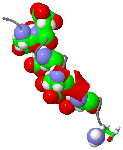
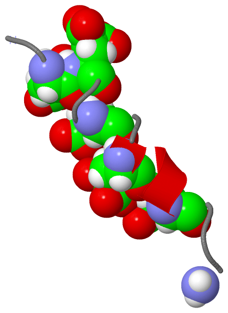
NMR StructureChain A from PDB Type:PROTEIN Length:18 aligned with CKG_CONGE | P07231 from UniProtKB/Swiss-Prot Length:100 Alignment length:18 90 CKG_CONGE 81 GEEELQENQELIREKSNG 98 SCOP domains d1awya_ A: SCOP domains CATH domains ------------------ CATH domains Pfam domains ------------------ Pfam domains SAPs(SNPs) ----V------------- SAPs(SNPs) PROSITE CONANTOKIN ---- PROSITE Transcript ------------------ Transcript 1awy A 1 GEeeLQeNQeLIReKSNx 18 || | 10 | | || | | | | 3-CGU | | | 4-CGU | | | 7-CGU | | 10-CGU | 14-CGU 18-NH2
|
||||||||||||||||||||
SCOP Domains (1, 1)
NMR Structure
 |
CATH Domains (0, 0)| (no "CATH Domain" information available for 1AWY) |
Pfam Domains (0, 0)| (no "Pfam Domain" information available for 1AWY) |
Gene Ontology (4, 4)|
NMR Structure(hide GO term definitions) Chain A (CKG_CONGE | P07231)
|
||||||||||||||||||||||||||||||||||||||||||
Interactive Views
|
||||||||||||||||||||||||||||||||||||||||||||||||||||||||||||||||||||||||||||||||||||||||||||||||||||||||||||||||||||||||||||||||||||
Still Images
|
||||||||||||||||
Databases
|
||||||||||||||||||||||||||||||||||||||||||||||||||||||||||||||||||||||||||||||||||||||||||||||||||||||||||||||||||||||||||||||||||||||||||||||||||||||||||||||||
Analysis Tools
|
|||||||||||||||||||||||||||||||||||||||||||||||||||||||||||||
Entries Sharing at Least One Protein Chain (UniProt ID)
Related Entries Specified in the PDB File
|
|