|
|
|
|
 Description
|
|
Compounds
|
||||||||||||||||||||||||||||
Chains, Units
Summary Information (see also Sequences/Alignments below) |
Ligands, Modified Residues, Ions (4, 5)| Asymmetric/Biological Unit (4, 5) |
Sites (6, 6)
Asymmetric Unit (6, 6)
|
SS Bonds (0, 0)| (no "SS Bond" information available for 1ATG) |
Cis Peptide Bonds (0, 0)| (no "Cis Peptide Bond" information available for 1ATG) |
SAPs(SNPs)/Variants (0, 0)| (no "SAP(SNP)/Variant" information available for 1ATG) |
PROSITE Motifs (0, 0)| (no "PROSITE Motif" information available for 1ATG) |
Exons (0, 0)| (no "Exon" information available for 1ATG) |
Sequences/Alignments
Asymmetric/Biological UnitChain A from PDB Type:PROTEIN Length:231 aligned with Q7SIH2_AZOVI | Q7SIH2 from UniProtKB/TrEMBL Length:231 Alignment length:231 10 20 30 40 50 60 70 80 90 100 110 120 130 140 150 160 170 180 190 200 210 220 230 Q7SIH2_AZOVI 1 ELKVVTATNFLGTLEQLAGQFAKQTGHAVVISSGSSGPVYAQIVNGAPYNVFFSADEKSPEKLDNQGFALPGSRFTYAIGKLVLWSAKPGLVDNQGKVLAGNGWRHIAISNPQIAPYGLAGTQVLTHLGLLDKLTAQERIVEANSVGQAHSQTASGAADLGFVALAQIIQAAAKIPGSHWFPPANYYEPIVQQAVITKSTAEKANAEQFMSWMKGPKAVAIIKAAGYVLPQ 231 SCOP domains d1atga_ A: Molybdate-binding protein, ModA SCOP domains CATH domains 1atgA01 A:2-81,A:191-232 Periplasmic binding protein-like II 1atgA02 A:82-190 Periplasmic binding protein-like II 1atgA01 A:2-81,A:191-232 CATH domains Pfam domains --------------------------------------------------------------------------------------------------------------------------------------------------------------------------------------------------------------------------------------- Pfam domains SAPs(SNPs) --------------------------------------------------------------------------------------------------------------------------------------------------------------------------------------------------------------------------------------- SAPs(SNPs) PROSITE --------------------------------------------------------------------------------------------------------------------------------------------------------------------------------------------------------------------------------------- PROSITE Transcript --------------------------------------------------------------------------------------------------------------------------------------------------------------------------------------------------------------------------------------- Transcript 1atg A 2 ELKVVTATNFLGTLEQLAGQFAKQTGHAVVISSGSSGPVYAQIVNGAPYNVFFSADEKSPEKLDNQGFALPGSRFTYAIGKLVLWSAKPGLVDNQGKVLAGNGWRHIAISNPQIAPYGLAGTQVLTHLGLLDKLTAQERIVEANSVGQAHSQTASGAADLGFVALAQIIQAAAKIPGSHWFPPANYYEPIVQQAVITKSTAEKANAEQFMSWMKGPKAVAIIKAAGYVLPQ 232 11 21 31 41 51 61 71 81 91 101 111 121 131 141 151 161 171 181 191 201 211 221 231
|
||||||||||||||||||||
SCOP Domains (1, 1)
Asymmetric/Biological Unit
 |
CATH Domains (1, 2)
Asymmetric/Biological Unit
|
Pfam Domains (0, 0)| (no "Pfam Domain" information available for 1ATG) |
Gene Ontology (3, 3)|
Asymmetric/Biological Unit(hide GO term definitions) Chain A (Q7SIH2_AZOVI | Q7SIH2)
|
||||||||||||||||||||||||||||||||||||
Interactive Views
|
||||||||||||||||||||||||||||||||||||||||||||||||||||||||||||||||||||||||||||||||||||||||||||||||||||||||||||||||||||||||||||||||||||||||||||||||||||||||||||||||||||||||||||||
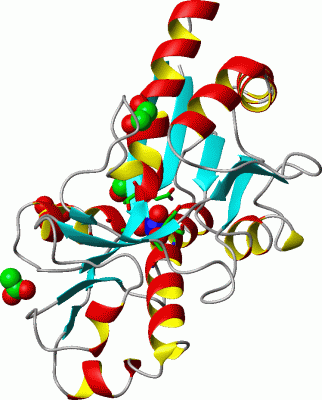
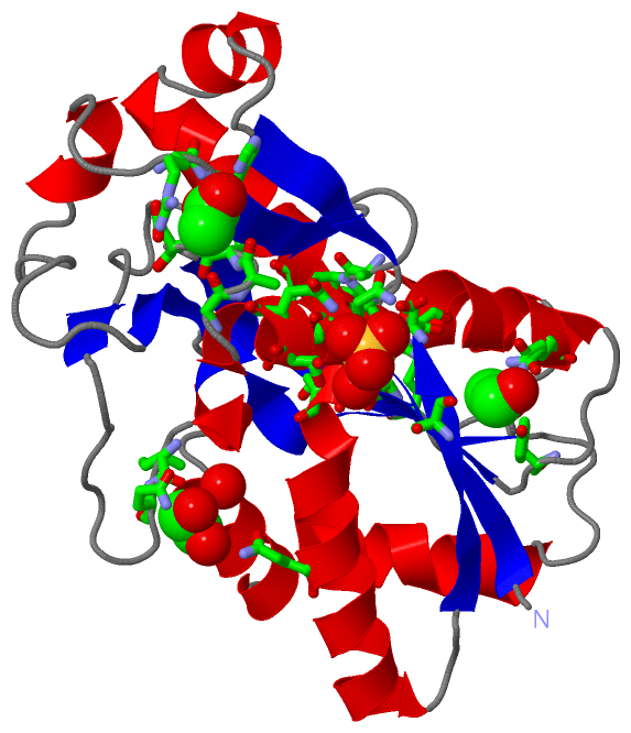
Still Images
|
||||||||||||||||
Databases
|
||||||||||||||||||||||||||||||||||||||||||||||||||||||||||||||||||||||||||||||||||||||||||||||||||||||||||||||||||||||||||||||||||||||||||||||||||||||||||||||||
Analysis Tools
|
|||||||||||||||||||||||||||||||||||||||||||||||||||||||||||||
Entries Sharing at Least One Protein Chain (UniProt ID)
Related Entries Specified in the PDB File
|
|