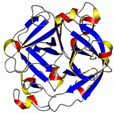
Asymmetric/Biological Unit
Chain A from PDB  Type:PROTEIN  Length:263
 aligned with API_ACHLY | P15636 from UniProtKB/Swiss-Prot  Length:653

    Alignment length:263
                                   215       225       235       245       255       265       275       285       295       305       315       325       335       345       355       365       375       385       395       405       415       425       435       445       455       465
            API_ACHLY   206 GVSGSCNIDVVCPEGDGRRDIIRAVGAYSKSGTLACTGSLVNNTANDRKMYFLTAHHCGMGTASTAASIVVYWNYQNSTCRAPNTPASGANGDGSMSQTQSGSTVKATYATSDFTLLELNNAANPAFNLFWAGWDRRDQNYPGAIAIHHPNVAEKRISNSTSPTSFVAWGGGAGTTHLNVQWQPSGGVTEPGSSGSPIYSPEKRVLGQLHGGPSSCSATGTNRSDQYGRVFTSWTGGGAAASRLSDWLDPASTGAQFIDGLDS 468
               SCOP domains d1arba_ A: Achromobacter protease                                                                                                                                                                                                                                       SCOP domains
               CATH domains 1arbA01 A:1-24,A:134-2311arbA02 A:25-131,A:233-262 Trypsin-like serine proteases                                                   --1arbA01 A:1-24,A:134-231 Trypsin-like serine proteases                                            -1arbA02 A:25-131,A:233-262    - CATH domains
               Pfam domains ----------------------------------------------------------------------------------------------------------------------------------------------------------------------------------------------------------------------------------------------------------------------- Pfam domains
         Sec.struct. author .......ee..hhhhh...hhhh.eeeeee..eeeeeeee..........eeeee.hhh..hhhhhhh.eee............hhhhhh........eee.eeeeeee....eeeeee....hhhhh.ee..ee........eeeeehhhhh..eeeee....eee.........eeeee...............eee.....eeeeeee.......hhhh.eeeeeehhhhhhh..hhhhhhhhhhh.......ee..ee. Sec.struct. author
                 SAPs(SNPs) ----------------------------------------------------------------------------------------------------------------------------------------------------------------------------------------------------------------------------------------------------------------------- SAPs(SNPs)
                    PROSITE ----------------------------------------------------------------------------------------------------------------------------------------------------------------------------------------------------------------------------------------------------------------------- PROSITE
                 Transcript ----------------------------------------------------------------------------------------------------------------------------------------------------------------------------------------------------------------------------------------------------------------------- Transcript
                 1arb A   1 GVSGSCNIDVVCPEGDGRRDIIRAVGAYSKSGTLACTGSLVNNTANDRKMYFLTAHHCGMGTASTAASIVVYWNYQNSTCRAPNTPASGANGDGSMSQTQSGSTVKATYATSDFTLLELNNAANPAFNLFWAGWDRRDQNYPGAIAIHHPNVAEKRISNSTSPTSFVAWGGGAGTTHLNVQWQPSGGVTEPGSSGSPIYSPEKRVLGQLHGGPSSCSATGTNRSDQYGRVFTSWTGGGAAASRLSDWLDPASTGAQFIDGLDS 263
                                    10        20        30        40        50        60        70        80        90       100       110       120       130       140       150       160       170       180       190       200       210       220       230       240       250       260
| Legend: |
|
→ Mismatch |
(orange background) |
| |
- |
→ Gap |
(green background, '-', border residues have a numbering label) |
| |
|
→ Modified Residue |
(blue background, lower-case, 'x' indicates undefined single-letter code, labelled with number + name) |
| |
x |
→ Chemical Group |
(purple background, 'x', labelled with number + name, e.g. ACE or NH2) |
| |
extra numbering lines below/above indicate numbering irregularities and modified residue names etc., number ends below/above '|' |
|
 Description